

2 Fundamentals of Retinal Optical Coherence Tomography
35
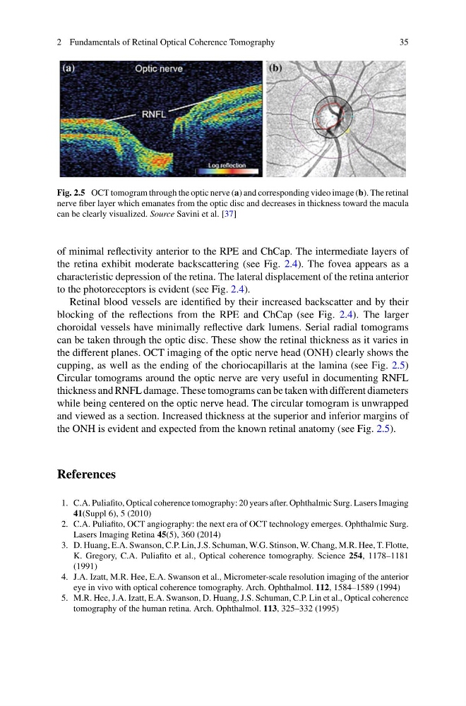
Fig. 2.5 OCT tomogram through the optic nerve (a) and corresponding video image (b). The retinal
nerve fiber layer which emanates from the optic disc and decreases in thickness toward the macula
can be clearly visualized. Source Savini et al. [37]
of minimal reflectivity anterior to the RPE and ChCap. The intermediate layers of
the retina exhibit moderate backscattering (see Fig. 2.4). The fovea appears as a
characteristic depression of the retina. The lateral displacement of the retina anterior
to the photoreceptors is evident (see Fig. 2.4).
Retinal blood vessels are identified by their increased backscatter and by their
blocking of the reflections from the RPE and ChCap (see Fig. 2.4). The larger
choroidal vessels have minimally reflective dark lumens. Serial radial tomograms
can be taken through the optic disc. These show the retinal thickness as it varies in
the different planes. OCT imaging of the optic nerve head (ONH) clearly shows the
cupping, as well as the ending of the choriocapillaris at the lamina (see Fig. 2.5)
Circular tomograms around the optic nerve are very useful in documenting RNFL
thickness and RNFL damage. These tomograms can be taken with different diameters
while being centered on the optic nerve head. The circular tomogram is unwrapped
and viewed as a section. Increased thickness at the superior and inferior margins of
the ONH is evident and expected from the known retinal anatomy (see Fig. 2.5).
References
1. C.A. Puliafito, Optical coherence tomography: 20 years after. Ophthalmic Surg. Lasers Imaging
41(Suppl 6), 5 (2010)
2. C.A. Puliafito, OCT angiography: the next era of OCT technology emerges. Ophthalmic Surg.
Lasers Imaging Retina 45(5), 360 (2014)
3. D. Huang, E.A. Swanson, C.P. Lin, J.S. Schuman, W.G. Stinson, W. Chang, M.R. Hee, T. Flotte,
K. Gregory, C.A. Puliafito et al., Optical coherence tomography. Science 254, 1178–1181
(1991)
4. J.A. Izatt, M.R. Hee, E.A. Swanson et al., Micrometer-scale resolution imaging of the anterior
eye in vivo with optical coherence tomography. Arch. Ophthalmol. 112, 1584–1589 (1994)
5. M.R. Hee, J.A. Izatt, E.A. Swanson, D. Huang, J.S. Schuman, C.P. Lin et al., Optical coherence
tomography of the human retina. Arch. Ophthalmol. 113, 325–332 (1995)
35
Fig. 2.5 OCT tomogram through the optic nerve (a) and corresponding video image (b). The retinal
nerve fiber layer which emanates from the optic disc and decreases in thickness toward the macula
can be clearly visualized. Source Savini et al. [37]
of minimal reflectivity anterior to the RPE and ChCap. The intermediate layers of
the retina exhibit moderate backscattering (see Fig. 2.4). The fovea appears as a
characteristic depression of the retina. The lateral displacement of the retina anterior
to the photoreceptors is evident (see Fig. 2.4).
Retinal blood vessels are identified by their increased backscatter and by their
blocking of the reflections from the RPE and ChCap (see Fig. 2.4). The larger
choroidal vessels have minimally reflective dark lumens. Serial radial tomograms
can be taken through the optic disc. These show the retinal thickness as it varies in
the different planes. OCT imaging of the optic nerve head (ONH) clearly shows the
cupping, as well as the ending of the choriocapillaris at the lamina (see Fig. 2.5)
Circular tomograms around the optic nerve are very useful in documenting RNFL
thickness and RNFL damage. These tomograms can be taken with different diameters
while being centered on the optic nerve head. The circular tomogram is unwrapped
and viewed as a section. Increased thickness at the superior and inferior margins of
the ONH is evident and expected from the known retinal anatomy (see Fig. 2.5).
References
1. C.A. Puliafito, Optical coherence tomography: 20 years after. Ophthalmic Surg. Lasers Imaging
41(Suppl 6), 5 (2010)
2. C.A. Puliafito, OCT angiography: the next era of OCT technology emerges. Ophthalmic Surg.
Lasers Imaging Retina 45(5), 360 (2014)
3. D. Huang, E.A. Swanson, C.P. Lin, J.S. Schuman, W.G. Stinson, W. Chang, M.R. Hee, T. Flotte,
K. Gregory, C.A. Puliafito et al., Optical coherence tomography. Science 254, 1178–1181
(1991)
4. J.A. Izatt, M.R. Hee, E.A. Swanson et al., Micrometer-scale resolution imaging of the anterior
eye in vivo with optical coherence tomography. Arch. Ophthalmol. 112, 1584–1589 (1994)
5. M.R. Hee, J.A. Izatt, E.A. Swanson, D. Huang, J.S. Schuman, C.P. Lin et al., Optical coherence
tomography of the human retina. Arch. Ophthalmol. 113, 325–332 (1995)