

2 Fundamentals of Retinal Optical Coherence Tomography
31
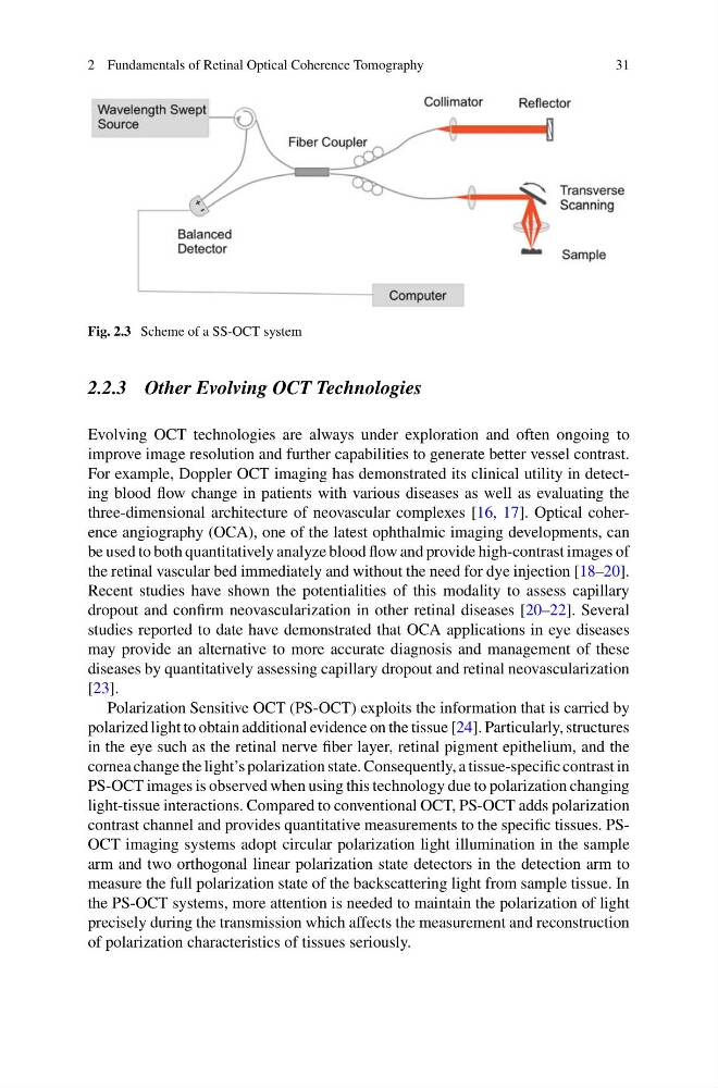
Fig. 2.3 Scheme of a SS-OCT system
2.2.3 Other Evolving OCT Technologies
Evolving OCT technologies are always under exploration and often ongoing to
improve image resolution and further capabilities to generate better vessel contrast.
For example, Doppler OCT imaging has demonstrated its clinical utility in detecting blood flow change in patients with various diseases as well as evaluating the
three-dimensional architecture of neovascular complexes [16, 17]. Optical coherence angiography (OCA), one of the latest ophthalmic imaging developments, can
be used to both quantitatively analyze blood flow and provide high-contrast images of
the retinal vascular bed immediately and without the need for dye injection [18–20].
Recent studies have shown the potentialities of this modality to assess capillary
dropout and confirm neovascularization in other retinal diseases [20–22]. Several
studies reported to date have demonstrated that OCA applications in eye diseases
may provide an alternative to more accurate diagnosis and management of these
diseases by quantitatively assessing capillary dropout and retinal neovascularization
[23].
Polarization Sensitive OCT (PS-OCT) exploits the information that is carried by
polarized light to obtain additional evidence on the tissue [24]. Particularly, structures
in the eye such as the retinal nerve fiber layer, retinal pigment epithelium, and the
cornea change the light’s polarization state. Consequently, a tissue-specific contrast in
PS-OCT images is observed when using this technology due to polarization changing
light-tissue interactions. Compared to conventional OCT, PS-OCT adds polarization
contrast channel and provides quantitative measurements to the specific tissues. PSOCT imaging systems adopt circular polarization light illumination in the sample
arm and two orthogonal linear polarization state detectors in the detection arm to
measure the full polarization state of the backscattering light from sample tissue. In
the PS-OCT systems, more attention is needed to maintain the polarization of light
precisely during the transmission which affects the measurement and reconstruction
of polarization characteristics of tissues seriously.
31
Fig. 2.3 Scheme of a SS-OCT system
2.2.3 Other Evolving OCT Technologies
Evolving OCT technologies are always under exploration and often ongoing to
improve image resolution and further capabilities to generate better vessel contrast.
For example, Doppler OCT imaging has demonstrated its clinical utility in detecting blood flow change in patients with various diseases as well as evaluating the
three-dimensional architecture of neovascular complexes [16, 17]. Optical coherence angiography (OCA), one of the latest ophthalmic imaging developments, can
be used to both quantitatively analyze blood flow and provide high-contrast images of
the retinal vascular bed immediately and without the need for dye injection [18–20].
Recent studies have shown the potentialities of this modality to assess capillary
dropout and confirm neovascularization in other retinal diseases [20–22]. Several
studies reported to date have demonstrated that OCA applications in eye diseases
may provide an alternative to more accurate diagnosis and management of these
diseases by quantitatively assessing capillary dropout and retinal neovascularization
[23].
Polarization Sensitive OCT (PS-OCT) exploits the information that is carried by
polarized light to obtain additional evidence on the tissue [24]. Particularly, structures
in the eye such as the retinal nerve fiber layer, retinal pigment epithelium, and the
cornea change the light’s polarization state. Consequently, a tissue-specific contrast in
PS-OCT images is observed when using this technology due to polarization changing
light-tissue interactions. Compared to conventional OCT, PS-OCT adds polarization
contrast channel and provides quantitative measurements to the specific tissues. PSOCT imaging systems adopt circular polarization light illumination in the sample
arm and two orthogonal linear polarization state detectors in the detection arm to
measure the full polarization state of the backscattering light from sample tissue. In
the PS-OCT systems, more attention is needed to maintain the polarization of light
precisely during the transmission which affects the measurement and reconstruction
of polarization characteristics of tissues seriously.