

30
D. Cabrera DeBuc et al.
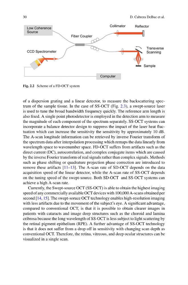
Fig. 2.2 Scheme of a FD-OCT system
of a dispersion grating and a linear detector, to measure the backscattering spectrum of the sample tissue. In the case of SS-OCT (Fig. 2.3), a swept-source laser
is used to tune the broad bandwidth frequency quickly. The reference arm length is
also fixed. A single point photodetector is employed in the detection arm to measure
the magnitude of each component of the spectrum separately. SS-OCT systems can
incorporate a balance detector design to suppress the impact of the laser heat fluctuation which can increase the sensitivity the sensitivity by approximately 10 dB.
The A-scan longitude information can be retrieved by inverse Fourier transform of
the spectrum data after interpolation processing which remaps the data linearly from
wavelength space to wavenumber space. FD-OCT suffers from artifacts such as the
direct current (DC), autocorrelation, and complex conjugate items which are caused
by the inverse Fourier transform of real signals rather than complex signals. Methods
such as phase shifting or quadrature projection phase correction are introduced to
remove these artifacts [11–13]. The A-scan rate of SD-OCT depends on the data
acquisition speed of the linear detector, while the A-scan rate of SS-OCT depends
on the tuning speed of the swept-source. Both SD-OCT and SS-OCT systems can
achieve a high A-scan rate.
Currently, the Swept-source OCT (SS-OCT) is able to obtain the highest imaging
speed of any commercially available OCT devices with 100,000 A-scans obtained per
second [14, 15]. The swept-soruce OCT technology enables high-resolution imaging
with less artifacts due to the movement of the subject’s eye. A significant advantage,
compared to conventional OCT, is that it is possible to obtain clearer images in
patients with cataracts and image deep structures such as the choroid and lamina
cribrosa because the long wavelength of SS-OCT is less subject to light scattering by
the retinal pigment epithelium (RPE). A further advantage of SS-OCT technology
is that it does not suffer from a drop off in sensitivity with changing scan depth as
conventional OCT. Therefore, the retina, vitreous, and deep ocular structures can be
visualized in a single scan.
D. Cabrera DeBuc et al.
Fig. 2.2 Scheme of a FD-OCT system
of a dispersion grating and a linear detector, to measure the backscattering spectrum of the sample tissue. In the case of SS-OCT (Fig. 2.3), a swept-source laser
is used to tune the broad bandwidth frequency quickly. The reference arm length is
also fixed. A single point photodetector is employed in the detection arm to measure
the magnitude of each component of the spectrum separately. SS-OCT systems can
incorporate a balance detector design to suppress the impact of the laser heat fluctuation which can increase the sensitivity the sensitivity by approximately 10 dB.
The A-scan longitude information can be retrieved by inverse Fourier transform of
the spectrum data after interpolation processing which remaps the data linearly from
wavelength space to wavenumber space. FD-OCT suffers from artifacts such as the
direct current (DC), autocorrelation, and complex conjugate items which are caused
by the inverse Fourier transform of real signals rather than complex signals. Methods
such as phase shifting or quadrature projection phase correction are introduced to
remove these artifacts [11–13]. The A-scan rate of SD-OCT depends on the data
acquisition speed of the linear detector, while the A-scan rate of SS-OCT depends
on the tuning speed of the swept-source. Both SD-OCT and SS-OCT systems can
achieve a high A-scan rate.
Currently, the Swept-source OCT (SS-OCT) is able to obtain the highest imaging
speed of any commercially available OCT devices with 100,000 A-scans obtained per
second [14, 15]. The swept-soruce OCT technology enables high-resolution imaging
with less artifacts due to the movement of the subject’s eye. A significant advantage,
compared to conventional OCT, is that it is possible to obtain clearer images in
patients with cataracts and image deep structures such as the choroid and lamina
cribrosa because the long wavelength of SS-OCT is less subject to light scattering by
the retinal pigment epithelium (RPE). A further advantage of SS-OCT technology
is that it does not suffer from a drop off in sensitivity with changing scan depth as
conventional OCT. Therefore, the retina, vitreous, and deep ocular structures can be
visualized in a single scan.