

13 Modeling and Prediction of Choroidal Neovascularization …
371
13.2.3 Preprocessing
In this longitudinal study, the images are collected one month each, and thus displacement of the retina in OCT images caused by different eye position during scanning is
inevitable. Therefore, to guarantee of prediction accuracy, it is important to registrate
images so that the change of lesion area at the same positions can be measured. The
first image is set as the reference image and the other images are registered to it using
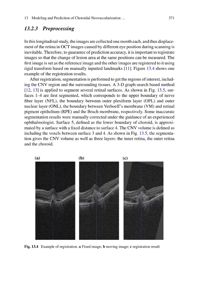
rigid transform based on manually inputted landmarks [11]. Figure 13.4 shows one
example of the registration results.
After registration, segmentation is performed to get the regions of interest, including the CNV region and the surrounding tissues. A 3-D graph-search based method
[12, 13] is applied to segment several retinal surfaces. As shown in Fig. 13.5, surfaces 1–4 are first segmented, which corresponds to the upper boundary of nerve
fiber layer (NFL), the boundary between outer plexiform layer (OPL) and outer
nuclear layer (ONL), the boundary between Verhoeff’s membrane (VM) and retinal
pigment epithelium (RPE) and the Bruch membrane, respectively. Some inaccurate
segmentation results were manually corrected under the guidance of an experienced
ophthalmologist. Surface 5, defined as the lower boundary of choroid, is approximated by a surface with a fixed distance to surface 4. The CNV volume is defined as
including the voxels between surface 3 and 4. As shown in Fig. 13.5, the segmentation gives the CNV volume as well as three layers: the inner retina, the outer retina
and the choroid.
Fig. 13.4 Example of registration. a Fixed image; b moving image; c registration result
371
13.2.3 Preprocessing
In this longitudinal study, the images are collected one month each, and thus displacement of the retina in OCT images caused by different eye position during scanning is
inevitable. Therefore, to guarantee of prediction accuracy, it is important to registrate
images so that the change of lesion area at the same positions can be measured. The
first image is set as the reference image and the other images are registered to it using
rigid transform based on manually inputted landmarks [11]. Figure 13.4 shows one
example of the registration results.
After registration, segmentation is performed to get the regions of interest, including the CNV region and the surrounding tissues. A 3-D graph-search based method
[12, 13] is applied to segment several retinal surfaces. As shown in Fig. 13.5, surfaces 1–4 are first segmented, which corresponds to the upper boundary of nerve
fiber layer (NFL), the boundary between outer plexiform layer (OPL) and outer
nuclear layer (ONL), the boundary between Verhoeff’s membrane (VM) and retinal
pigment epithelium (RPE) and the Bruch membrane, respectively. Some inaccurate
segmentation results were manually corrected under the guidance of an experienced
ophthalmologist. Surface 5, defined as the lower boundary of choroid, is approximated by a surface with a fixed distance to surface 4. The CNV volume is defined as
including the voxels between surface 3 and 4. As shown in Fig. 13.5, the segmentation gives the CNV volume as well as three layers: the inner retina, the outer retina
and the choroid.
Fig. 13.4 Example of registration. a Fixed image; b moving image; c registration result