

362
L. Pan and X. Chen
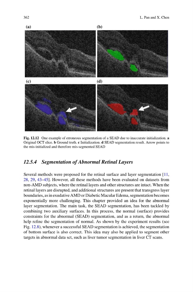
Fig. 12.12 One example of erroneous segmentation of a SEAD due to inaccurate initialization. a
Original OCT slice. b Ground truth. c Initialization. d SEAD segmentation result. Arrow points to
the mis-initialized and therefore mis-segmented SEAD
12.5.4 Segmentation of Abnormal Retinal Layers
Several methods were proposed for the retinal surface and layer segmentation [11,
28, 29, 43–45]. However, all these methods have been evaluated on datasets from
non-AMD subjects, where the retinal layers and other structures are intact. When the
retinal layers are disrupted, and additional structures are present that transgress layer
boundaries, as in exudative AMD or Diabetic Macular Edema, segmentation becomes
exponentially more challenging. This chapter provided an idea for the abnormal
layer segmentation. The main task, the SEAD segmentation, has been tackled by
combining two auxiliary surfaces. In this process, the normal (surface) provides
constraints for the abnormal (SEAD) segmentation, and as a return, the abnormal
help refine the segmentation of normal. As shown by the experiment results (see
Fig. 12.8), whenever a successful SEAD segmentation is achieved, the segmentation
of bottom surface is also correct. This idea may also be applied to segment other
targets in abnormal data set, such as liver tumor segmentation in liver CT scans.
L. Pan and X. Chen
Fig. 12.12 One example of erroneous segmentation of a SEAD due to inaccurate initialization. a
Original OCT slice. b Ground truth. c Initialization. d SEAD segmentation result. Arrow points to
the mis-initialized and therefore mis-segmented SEAD
12.5.4 Segmentation of Abnormal Retinal Layers
Several methods were proposed for the retinal surface and layer segmentation [11,
28, 29, 43–45]. However, all these methods have been evaluated on datasets from
non-AMD subjects, where the retinal layers and other structures are intact. When the
retinal layers are disrupted, and additional structures are present that transgress layer
boundaries, as in exudative AMD or Diabetic Macular Edema, segmentation becomes
exponentially more challenging. This chapter provided an idea for the abnormal
layer segmentation. The main task, the SEAD segmentation, has been tackled by
combining two auxiliary surfaces. In this process, the normal (surface) provides
constraints for the abnormal (SEAD) segmentation, and as a return, the abnormal
help refine the segmentation of normal. As shown by the experiment results (see
Fig. 12.8), whenever a successful SEAD segmentation is achieved, the segmentation
of bottom surface is also correct. This idea may also be applied to segment other
targets in abnormal data set, such as liver tumor segmentation in liver CT scans.