

12 Segmentation of Symptomatic Exudate-Associated …
349
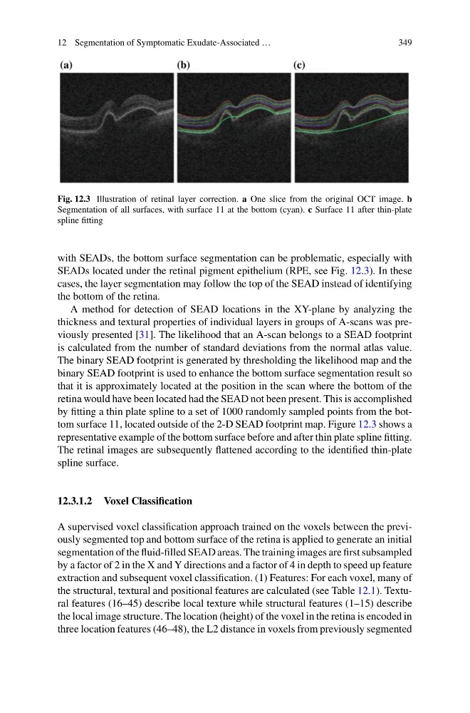
Fig. 12.3 Illustration of retinal layer correction. a One slice from the original OCT image. b
Segmentation of all surfaces, with surface 11 at the bottom (cyan). c Surface 11 after thin-plate
spline fitting
with SEADs, the bottom surface segmentation can be problematic, especially with
SEADs located under the retinal pigment epithelium (RPE, see Fig. 12.3). In these
cases, the layer segmentation may follow the top of the SEAD instead of identifying
the bottom of the retina.
A method for detection of SEAD locations in the XY-plane by analyzing the
thickness and textural properties of individual layers in groups of A-scans was previously presented [31]. The likelihood that an A-scan belongs to a SEAD footprint
is calculated from the number of standard deviations from the normal atlas value.
The binary SEAD footprint is generated by thresholding the likelihood map and the
binary SEAD footprint is used to enhance the bottom surface segmentation result so
that it is approximately located at the position in the scan where the bottom of the
retina would have been located had the SEAD not been present. This is accomplished
by fitting a thin plate spline to a set of 1000 randomly sampled points from the bottom surface 11, located outside of the 2-D SEAD footprint map. Figure 12.3 shows a
representative example of the bottom surface before and after thin plate spline fitting.
The retinal images are subsequently flattened according to the identified thin-plate
spline surface.
12.3.1.2 Voxel Classification
A supervised voxel classification approach trained on the voxels between the previously segmented top and bottom surface of the retina is applied to generate an initial
segmentation of the fluid-filled SEAD areas. The training images are first subsampled
by a factor of 2 in the X and Y directions and a factor of 4 in depth to speed up feature
extraction and subsequent voxel classification. (1) Features: For each voxel, many of
the structural, textural and positional features are calculated (see Table 12.1). Textural features (16–45) describe local texture while structural features (1–15) describe
the local image structure. The location (height) of the voxel in the retina is encoded in
three location features (46–48), the L2 distance in voxels from previously segmented
349
Fig. 12.3 Illustration of retinal layer correction. a One slice from the original OCT image. b
Segmentation of all surfaces, with surface 11 at the bottom (cyan). c Surface 11 after thin-plate
spline fitting
with SEADs, the bottom surface segmentation can be problematic, especially with
SEADs located under the retinal pigment epithelium (RPE, see Fig. 12.3). In these
cases, the layer segmentation may follow the top of the SEAD instead of identifying
the bottom of the retina.
A method for detection of SEAD locations in the XY-plane by analyzing the
thickness and textural properties of individual layers in groups of A-scans was previously presented [31]. The likelihood that an A-scan belongs to a SEAD footprint
is calculated from the number of standard deviations from the normal atlas value.
The binary SEAD footprint is generated by thresholding the likelihood map and the
binary SEAD footprint is used to enhance the bottom surface segmentation result so
that it is approximately located at the position in the scan where the bottom of the
retina would have been located had the SEAD not been present. This is accomplished
by fitting a thin plate spline to a set of 1000 randomly sampled points from the bottom surface 11, located outside of the 2-D SEAD footprint map. Figure 12.3 shows a
representative example of the bottom surface before and after thin plate spline fitting.
The retinal images are subsequently flattened according to the identified thin-plate
spline surface.
12.3.1.2 Voxel Classification
A supervised voxel classification approach trained on the voxels between the previously segmented top and bottom surface of the retina is applied to generate an initial
segmentation of the fluid-filled SEAD areas. The training images are first subsampled
by a factor of 2 in the X and Y directions and a factor of 4 in depth to speed up feature
extraction and subsequent voxel classification. (1) Features: For each voxel, many of
the structural, textural and positional features are calculated (see Table 12.1). Textural features (16–45) describe local texture while structural features (1–15) describe
the local image structure. The location (height) of the voxel in the retina is encoded in
three location features (46–48), the L2 distance in voxels from previously segmented