

11 Segmentation and Visualization of Drusen …
305
Input
image
RPE layer
estimation
Generation of GA
projection image
GA segmentation based on geometric active contour model
Output
result
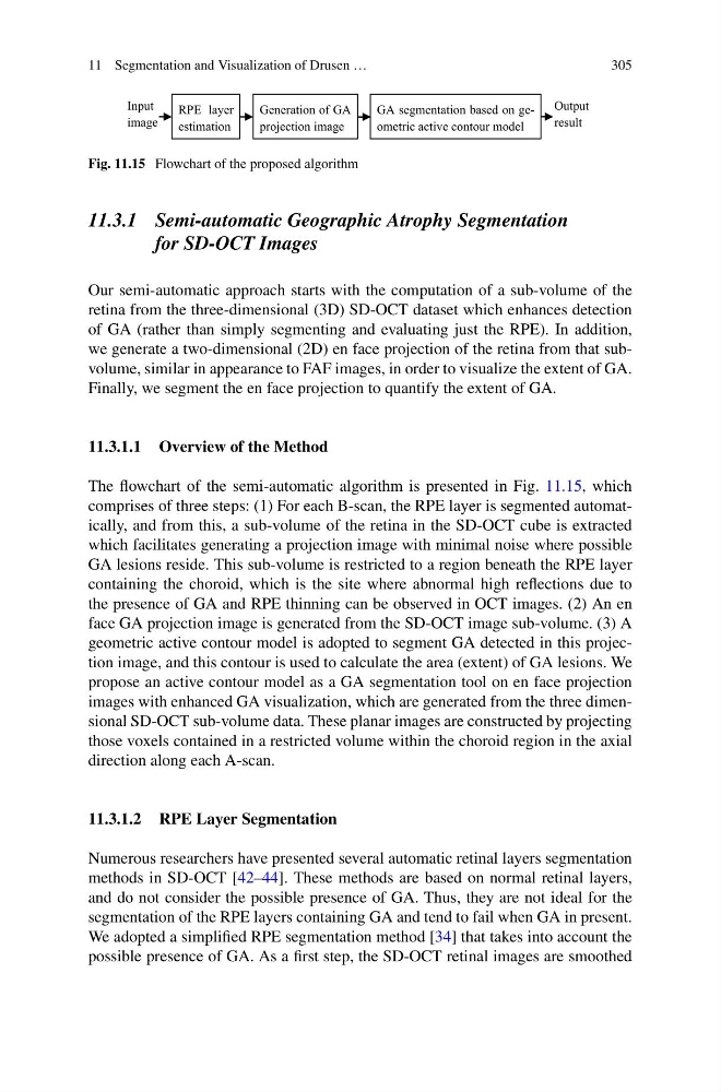
Fig. 11.15 Flowchart of the proposed algorithm
11.3.1 Semi-automatic Geographic Atrophy Segmentation
for SD-OCT Images
Our semi-automatic approach starts with the computation of a sub-volume of the
retina from the three-dimensional (3D) SD-OCT dataset which enhances detection
of GA (rather than simply segmenting and evaluating just the RPE). In addition,
we generate a two-dimensional (2D) en face projection of the retina from that subvolume, similar in appearance to FAF images, in order to visualize the extent of GA.
Finally, we segment the en face projection to quantify the extent of GA.
11.3.1.1 Overview of the Method
The flowchart of the semi-automatic algorithm is presented in Fig. 11.15, which
comprises of three steps: (1) For each B-scan, the RPE layer is segmented automatically, and from this, a sub-volume of the retina in the SD-OCT cube is extracted
which facilitates generating a projection image with minimal noise where possible
GA lesions reside. This sub-volume is restricted to a region beneath the RPE layer
containing the choroid, which is the site where abnormal high reflections due to
the presence of GA and RPE thinning can be observed in OCT images. (2) An en
face GA projection image is generated from the SD-OCT image sub-volume. (3) A
geometric active contour model is adopted to segment GA detected in this projection image, and this contour is used to calculate the area (extent) of GA lesions. We
propose an active contour model as a GA segmentation tool on en face projection
images with enhanced GA visualization, which are generated from the three dimensional SD-OCT sub-volume data. These planar images are constructed by projecting
those voxels contained in a restricted volume within the choroid region in the axial
direction along each A-scan.
11.3.1.2 RPE Layer Segmentation
Numerous researchers have presented several automatic retinal layers segmentation
methods in SD-OCT [42–44]. These methods are based on normal retinal layers,
and do not consider the possible presence of GA. Thus, they are not ideal for the
segmentation of the RPE layers containing GA and tend to fail when GA in present.
We adopted a simplified RPE segmentation method [34] that takes into account the
possible presence of GA. As a first step, the SD-OCT retinal images are smoothed
305
Input
image
RPE layer
estimation
Generation of GA
projection image
GA segmentation based on geometric active contour model
Output
result
Fig. 11.15 Flowchart of the proposed algorithm
11.3.1 Semi-automatic Geographic Atrophy Segmentation
for SD-OCT Images
Our semi-automatic approach starts with the computation of a sub-volume of the
retina from the three-dimensional (3D) SD-OCT dataset which enhances detection
of GA (rather than simply segmenting and evaluating just the RPE). In addition,
we generate a two-dimensional (2D) en face projection of the retina from that subvolume, similar in appearance to FAF images, in order to visualize the extent of GA.
Finally, we segment the en face projection to quantify the extent of GA.
11.3.1.1 Overview of the Method
The flowchart of the semi-automatic algorithm is presented in Fig. 11.15, which
comprises of three steps: (1) For each B-scan, the RPE layer is segmented automatically, and from this, a sub-volume of the retina in the SD-OCT cube is extracted
which facilitates generating a projection image with minimal noise where possible
GA lesions reside. This sub-volume is restricted to a region beneath the RPE layer
containing the choroid, which is the site where abnormal high reflections due to
the presence of GA and RPE thinning can be observed in OCT images. (2) An en
face GA projection image is generated from the SD-OCT image sub-volume. (3) A
geometric active contour model is adopted to segment GA detected in this projection image, and this contour is used to calculate the area (extent) of GA lesions. We
propose an active contour model as a GA segmentation tool on en face projection
images with enhanced GA visualization, which are generated from the three dimensional SD-OCT sub-volume data. These planar images are constructed by projecting
those voxels contained in a restricted volume within the choroid region in the axial
direction along each A-scan.
11.3.1.2 RPE Layer Segmentation
Numerous researchers have presented several automatic retinal layers segmentation
methods in SD-OCT [42–44]. These methods are based on normal retinal layers,
and do not consider the possible presence of GA. Thus, they are not ideal for the
segmentation of the RPE layers containing GA and tend to fail when GA in present.
We adopted a simplified RPE segmentation method [34] that takes into account the
possible presence of GA. As a first step, the SD-OCT retinal images are smoothed