

11 Segmentation and Visualization of Drusen …
285
Input
image
Image
denoising
RNFL
removal
RPE
segmentation
Drusen
segmentation
Drusen
projection
Elimination of false
positive drusen
Drusen refinement on
projection image image
Drusen
smoothing
Output
result
1
2
3
6
5
4
7
8
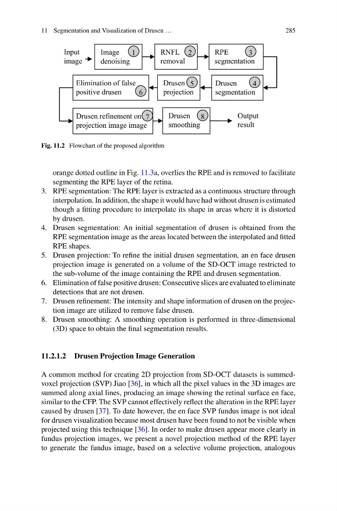
Fig. 11.2 Flowchart of the proposed algorithm
orange dotted outline in Fig. 11.3a, overlies the RPE and is removed to facilitate
segmenting the RPE layer of the retina.
3. RPE segmentation: The RPE layer is extracted as a continuous structure through
interpolation. In addition, the shape it would have had without drusen is estimated
though a fitting procedure to interpolate its shape in areas where it is distorted
by drusen.
4. Drusen segmentation: An initial segmentation of drusen is obtained from the
RPE segmentation image as the areas located between the interpolated and fitted
RPE shapes.
5. Drusen projection: To refine the initial drusen segmentation, an en face drusen
projection image is generated on a volume of the SD-OCT image restricted to
the sub-volume of the image containing the RPE and drusen segmentation.
6. Elimination of false positive drusen: Consecutive slices are evaluated to eliminate
detections that are not drusen.
7. Drusen refinement: The intensity and shape information of drusen on the projection image are utilized to remove false drusen.
8. Drusen smoothing: A smoothing operation is performed in three-dimensional
(3D) space to obtain the final segmentation results.
11.2.1.2 Drusen Projection Image Generation
A common method for creating 2D projection from SD-OCT datasets is summedvoxel projection (SVP) Jiao [36], in which all the pixel values in the 3D images are
summed along axial lines, producing an image showing the retinal surface en face,
similar to the CFP. The SVP cannot effectively reflect the alteration in the RPE layer
caused by drusen [37]. To date however, the en face SVP fundus image is not ideal
for drusen visualization because most drusen have been found to not be visible when
projected using this technique [36]. In order to make drusen appear more clearly in
fundus projection images, we present a novel projection method of the RPE layer
to generate the fundus image, based on a selective volume projection, analogous
285
Input
image
Image
denoising
RNFL
removal
RPE
segmentation
Drusen
segmentation
Drusen
projection
Elimination of false
positive drusen
Drusen refinement on
projection image image
Drusen
smoothing
Output
result
1
2
3
6
5
4
7
8
Fig. 11.2 Flowchart of the proposed algorithm
orange dotted outline in Fig. 11.3a, overlies the RPE and is removed to facilitate
segmenting the RPE layer of the retina.
3. RPE segmentation: The RPE layer is extracted as a continuous structure through
interpolation. In addition, the shape it would have had without drusen is estimated
though a fitting procedure to interpolate its shape in areas where it is distorted
by drusen.
4. Drusen segmentation: An initial segmentation of drusen is obtained from the
RPE segmentation image as the areas located between the interpolated and fitted
RPE shapes.
5. Drusen projection: To refine the initial drusen segmentation, an en face drusen
projection image is generated on a volume of the SD-OCT image restricted to
the sub-volume of the image containing the RPE and drusen segmentation.
6. Elimination of false positive drusen: Consecutive slices are evaluated to eliminate
detections that are not drusen.
7. Drusen refinement: The intensity and shape information of drusen on the projection image are utilized to remove false drusen.
8. Drusen smoothing: A smoothing operation is performed in three-dimensional
(3D) space to obtain the final segmentation results.
11.2.1.2 Drusen Projection Image Generation
A common method for creating 2D projection from SD-OCT datasets is summedvoxel projection (SVP) Jiao [36], in which all the pixel values in the 3D images are
summed along axial lines, producing an image showing the retinal surface en face,
similar to the CFP. The SVP cannot effectively reflect the alteration in the RPE layer
caused by drusen [37]. To date however, the en face SVP fundus image is not ideal
for drusen visualization because most drusen have been found to not be visible when
projected using this technique [36]. In order to make drusen appear more clearly in
fundus projection images, we present a novel projection method of the RPE layer
to generate the fundus image, based on a selective volume projection, analogous