

10 Layer Segmentation and Analysis for Retina with Diseases
257
Table 10.5 p-values of the proposed algorithm versus reference standards and the Iowa reference
algorithm [14] for B-scans with PED
Surface #
p value Algo. versus Ref.
p value Algo.versus [14]
1
0.001
0.4765
2
0.0733
0.001
4
0.0062
0.001
5
0.9503
0.0430
6
0.0056
0.0132
7
0.0981
0.0140
10
0.2834
0.001
11
0.5595
0.001
Overall
0.1806
0.001
Here numbers in bold indicate statistically significantly better performance
and 12 represent the same surface and their detection results will mostly overlap.
Obtained with a large smoothness constraint, surface 11 is likely to be less smooth
than surface 12 due to the impact of noise, but the regions between surfaces 11 and 12
will be excluded as false positives in PED detection. Then flattening with respect to
surface 11 is no more than a step which further removes the eye movement artifacts,
as did in [1, 2, 5]. Additionally, correction of surfaces 7–9 is not needed and this step
will be automatically skipped when no PED region is detected.
To test the performance in normal data, the method was applied to OCT images
from a control group of 20 normal subjects. Table 10.6 shows the mean and standard
deviation of unsigned border positioning errors for each surface, compared with
inter-observer variability and the errors resulting from employing the Iowa Reference
Algorithm [14]. The p-values are shown in Table 10.7, with bold fonts indicating that
the proposed method has statistically significantly better performance. The overall
mean unsigned error of the proposed algorithm is significantly smaller than the mean
unsigned difference between two observers. Compared with [14], the overall error
is statistically indistinguishable.
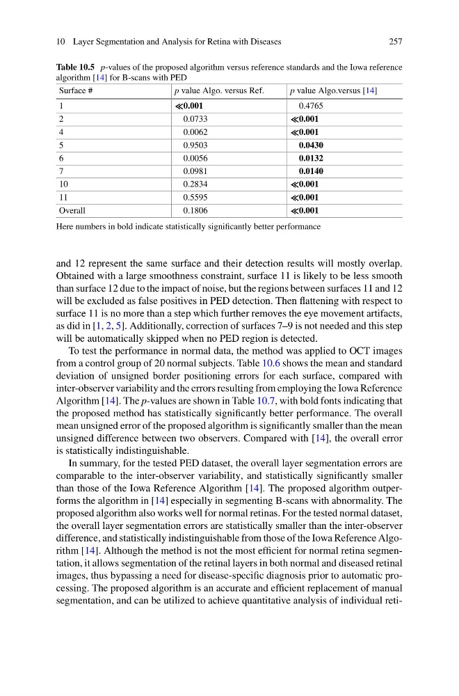
In summary, for the tested PED dataset, the overall layer segmentation errors are
comparable to the inter-observer variability, and statistically significantly smaller
than those of the Iowa Reference Algorithm [14]. The proposed algorithm outperforms the algorithm in [14] especially in segmenting B-scans with abnormality. The
proposed algorithm also works well for normal retinas. For the tested normal dataset,
the overall layer segmentation errors are statistically smaller than the inter-observer
difference, and statistically indistinguishable from those of the Iowa Reference Algorithm [14]. Although the method is not the most efficient for normal retina segmentation, it allows segmentation of the retinal layers in both normal and diseased retinal
images, thus bypassing a need for disease-specific diagnosis prior to automatic processing. The proposed algorithm is an accurate and efficient replacement of manual
segmentation, and can be utilized to achieve quantitative analysis of individual reti-
257
Table 10.5 p-values of the proposed algorithm versus reference standards and the Iowa reference
algorithm [14] for B-scans with PED
Surface #
p value Algo. versus Ref.
p value Algo.versus [14]
1
0.001
0.4765
2
0.0733
0.001
4
0.0062
0.001
5
0.9503
0.0430
6
0.0056
0.0132
7
0.0981
0.0140
10
0.2834
0.001
11
0.5595
0.001
Overall
0.1806
0.001
Here numbers in bold indicate statistically significantly better performance
and 12 represent the same surface and their detection results will mostly overlap.
Obtained with a large smoothness constraint, surface 11 is likely to be less smooth
than surface 12 due to the impact of noise, but the regions between surfaces 11 and 12
will be excluded as false positives in PED detection. Then flattening with respect to
surface 11 is no more than a step which further removes the eye movement artifacts,
as did in [1, 2, 5]. Additionally, correction of surfaces 7–9 is not needed and this step
will be automatically skipped when no PED region is detected.
To test the performance in normal data, the method was applied to OCT images
from a control group of 20 normal subjects. Table 10.6 shows the mean and standard
deviation of unsigned border positioning errors for each surface, compared with
inter-observer variability and the errors resulting from employing the Iowa Reference
Algorithm [14]. The p-values are shown in Table 10.7, with bold fonts indicating that
the proposed method has statistically significantly better performance. The overall
mean unsigned error of the proposed algorithm is significantly smaller than the mean
unsigned difference between two observers. Compared with [14], the overall error
is statistically indistinguishable.
In summary, for the tested PED dataset, the overall layer segmentation errors are
comparable to the inter-observer variability, and statistically significantly smaller
than those of the Iowa Reference Algorithm [14]. The proposed algorithm outperforms the algorithm in [14] especially in segmenting B-scans with abnormality. The
proposed algorithm also works well for normal retinas. For the tested normal dataset,
the overall layer segmentation errors are statistically smaller than the inter-observer
difference, and statistically indistinguishable from those of the Iowa Reference Algorithm [14]. Although the method is not the most efficient for normal retina segmentation, it allows segmentation of the retinal layers in both normal and diseased retinal
images, thus bypassing a need for disease-specific diagnosis prior to automatic processing. The proposed algorithm is an accurate and efficient replacement of manual
segmentation, and can be utilized to achieve quantitative analysis of individual reti-