

238
K. K. Vupparaboina et al.
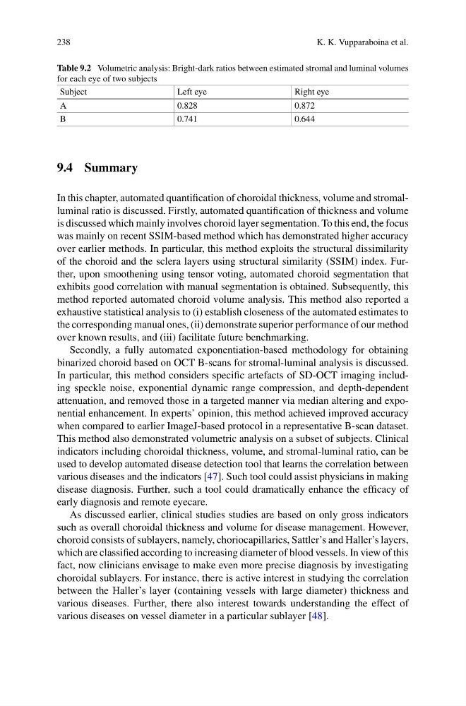
Table 9.2 Volumetric analysis: Bright-dark ratios between estimated stromal and luminal volumes
for each eye of two subjects
Subject
Left eye
Right eye
A
0.828
0.872
B
0.741
0.644
9.4 Summary
In this chapter, automated quantification of choroidal thickness, volume and stromalluminal ratio is discussed. Firstly, automated quantification of thickness and volume
is discussed which mainly involves choroid layer segmentation. To this end, the focus
was mainly on recent SSIM-based method which has demonstrated higher accuracy
over earlier methods. In particular, this method exploits the structural dissimilarity
of the choroid and the sclera layers using structural similarity (SSIM) index. Further, upon smoothening using tensor voting, automated choroid segmentation that
exhibits good correlation with manual segmentation is obtained. Subsequently, this
method reported automated choroid volume analysis. This method also reported a
exhaustive statistical analysis to (i) establish closeness of the automated estimates to
the corresponding manual ones, (ii) demonstrate superior performance of our method
over known results, and (iii) facilitate future benchmarking.
Secondly, a fully automated exponentiation-based methodology for obtaining
binarized choroid based on OCT B-scans for stromal-luminal analysis is discussed.
In particular, this method considers specific artefacts of SD-OCT imaging including speckle noise, exponential dynamic range compression, and depth-dependent
attenuation, and removed those in a targeted manner via median altering and exponential enhancement. In experts’ opinion, this method achieved improved accuracy
when compared to earlier ImageJ-based protocol in a representative B-scan dataset.
This method also demonstrated volumetric analysis on a subset of subjects. Clinical
indicators including choroidal thickness, volume, and stromal-luminal ratio, can be
used to develop automated disease detection tool that learns the correlation between
various diseases and the indicators [47]. Such tool could assist physicians in making
disease diagnosis. Further, such a tool could dramatically enhance the efficacy of
early diagnosis and remote eyecare.
As discussed earlier, clinical studies studies are based on only gross indicators
such as overall choroidal thickness and volume for disease management. However,
choroid consists of sublayers, namely, choriocapillaries, Sattler’s and Haller’s layers,
which are classified according to increasing diameter of blood vessels. In view of this
fact, now clinicians envisage to make even more precise diagnosis by investigating
choroidal sublayers. For instance, there is active interest in studying the correlation
between the Haller’s layer (containing vessels with large diameter) thickness and
various diseases. Further, there also interest towards understanding the effect of
various diseases on vessel diameter in a particular sublayer [48].
K. K. Vupparaboina et al.
Table 9.2 Volumetric analysis: Bright-dark ratios between estimated stromal and luminal volumes
for each eye of two subjects
Subject
Left eye
Right eye
A
0.828
0.872
B
0.741
0.644
9.4 Summary
In this chapter, automated quantification of choroidal thickness, volume and stromalluminal ratio is discussed. Firstly, automated quantification of thickness and volume
is discussed which mainly involves choroid layer segmentation. To this end, the focus
was mainly on recent SSIM-based method which has demonstrated higher accuracy
over earlier methods. In particular, this method exploits the structural dissimilarity
of the choroid and the sclera layers using structural similarity (SSIM) index. Further, upon smoothening using tensor voting, automated choroid segmentation that
exhibits good correlation with manual segmentation is obtained. Subsequently, this
method reported automated choroid volume analysis. This method also reported a
exhaustive statistical analysis to (i) establish closeness of the automated estimates to
the corresponding manual ones, (ii) demonstrate superior performance of our method
over known results, and (iii) facilitate future benchmarking.
Secondly, a fully automated exponentiation-based methodology for obtaining
binarized choroid based on OCT B-scans for stromal-luminal analysis is discussed.
In particular, this method considers specific artefacts of SD-OCT imaging including speckle noise, exponential dynamic range compression, and depth-dependent
attenuation, and removed those in a targeted manner via median altering and exponential enhancement. In experts’ opinion, this method achieved improved accuracy
when compared to earlier ImageJ-based protocol in a representative B-scan dataset.
This method also demonstrated volumetric analysis on a subset of subjects. Clinical
indicators including choroidal thickness, volume, and stromal-luminal ratio, can be
used to develop automated disease detection tool that learns the correlation between
various diseases and the indicators [47]. Such tool could assist physicians in making
disease diagnosis. Further, such a tool could dramatically enhance the efficacy of
early diagnosis and remote eyecare.
As discussed earlier, clinical studies studies are based on only gross indicators
such as overall choroidal thickness and volume for disease management. However,
choroid consists of sublayers, namely, choriocapillaries, Sattler’s and Haller’s layers,
which are classified according to increasing diameter of blood vessels. In view of this
fact, now clinicians envisage to make even more precise diagnosis by investigating
choroidal sublayers. For instance, there is active interest in studying the correlation
between the Haller’s layer (containing vessels with large diameter) thickness and
various diseases. Further, there also interest towards understanding the effect of
various diseases on vessel diameter in a particular sublayer [48].