

186
E. Gao et al.
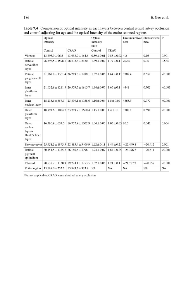
Table 7.4 Comparison of optical intensity in each layers between central retinal artery occlusion
and control adjusting for age and the optical intensity of the entire scanned regions
Optical
intensity
Optical
intensity
ratio
Unstandardized
beta
Standardized
beta
P
Control
CRAO
Control
CRAO
Vitreous
13,893.9 ± 96.5
13,953.9 ± 164.6 0.89 ± 0.01 0.88 ± 0.02 4.2
0.16
0.901
Retinal
nerve fiber
layer
26,598.5 ± 1598.1 28,232.6 ± 2120 1.69 ± 0.09 1.77 ± 0.11 202.6
0.05
0.584
Retinal
ganglion cell
layer
21,567.8 ± 1301.4 26,219.3 ± 1980.1 1.37 ± 0.06 1.64 ± 0.11 3709.4
0.657
<0.001
Inner
plexiform
layer
21,052.8 ± 1211.5 26,559.5 ± 1915.7 1.34 ± 0.06 1.66 ± 0.1 4441
0.702
<0.001
Inner
nuclear layer
18,235.6 ± 857.9 23,899.1 ± 1758.6 1.16 ± 0.04 1.5 ± 0.09 4863.3
0.777
<0.001
Outer
plexiform
layer
18,791.6 ± 1084.7 23,389.7 ± 1660.4 1.15 ± 0.03 1.4 ± 0.1 3708.8
0.694
<0.001
Outer
nuclear
layer +
Henle’s fiber
layer
16,380.9 ± 657.5 16,757.9 ± 1002.9 1.04 ± 0.03 1.05 ± 0.05 80.3
0.047
0.664
Photoreceptor 25,458.3 ± 1893.3 22,885.4 ± 3406.9 1.62 ± 0.11 1.44 ± 0.21 −22,440.8
−20.412
0.001
Retinal
pigment
epithelium
30,454.5 ± 1375.2 26,160.6 ± 3998 1.94 ± 0.07 1.64 ± 0.25 −24,376.7
−20.611
<0.001
Choroid
20,638.7 ± 1130.9 19,224.1 ± 1753.5 1.32 ± 0.06 1.21 ± 0.1 −21,787.7
−20.559
<0.001
Entire region 15,688.0 ± 252.7 15,943.2 ± 333.4 NA
NA
NA
NA
NA
NA: not applicable; CRAO: central retinal artery occlusion
E. Gao et al.
Table 7.4 Comparison of optical intensity in each layers between central retinal artery occlusion
and control adjusting for age and the optical intensity of the entire scanned regions
Optical
intensity
Optical
intensity
ratio
Unstandardized
beta
Standardized
beta
P
Control
CRAO
Control
CRAO
Vitreous
13,893.9 ± 96.5
13,953.9 ± 164.6 0.89 ± 0.01 0.88 ± 0.02 4.2
0.16
0.901
Retinal
nerve fiber
layer
26,598.5 ± 1598.1 28,232.6 ± 2120 1.69 ± 0.09 1.77 ± 0.11 202.6
0.05
0.584
Retinal
ganglion cell
layer
21,567.8 ± 1301.4 26,219.3 ± 1980.1 1.37 ± 0.06 1.64 ± 0.11 3709.4
0.657
<0.001
Inner
plexiform
layer
21,052.8 ± 1211.5 26,559.5 ± 1915.7 1.34 ± 0.06 1.66 ± 0.1 4441
0.702
<0.001
Inner
nuclear layer
18,235.6 ± 857.9 23,899.1 ± 1758.6 1.16 ± 0.04 1.5 ± 0.09 4863.3
0.777
<0.001
Outer
plexiform
layer
18,791.6 ± 1084.7 23,389.7 ± 1660.4 1.15 ± 0.03 1.4 ± 0.1 3708.8
0.694
<0.001
Outer
nuclear
layer +
Henle’s fiber
layer
16,380.9 ± 657.5 16,757.9 ± 1002.9 1.04 ± 0.03 1.05 ± 0.05 80.3
0.047
0.664
Photoreceptor 25,458.3 ± 1893.3 22,885.4 ± 3406.9 1.62 ± 0.11 1.44 ± 0.21 −22,440.8
−20.412
0.001
Retinal
pigment
epithelium
30,454.5 ± 1375.2 26,160.6 ± 3998 1.94 ± 0.07 1.64 ± 0.25 −24,376.7
−20.611
<0.001
Choroid
20,638.7 ± 1130.9 19,224.1 ± 1753.5 1.32 ± 0.06 1.21 ± 0.1 −21,787.7
−20.559
<0.001
Entire region 15,688.0 ± 252.7 15,943.2 ± 333.4 NA
NA
NA
NA
NA
NA: not applicable; CRAO: central retinal artery occlusion