

184
E. Gao et al.
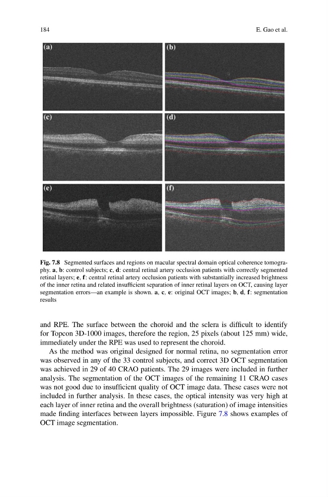
Fig. 7.8 Segmented surfaces and regions on macular spectral domain optical coherence tomography. a, b: control subjects; c, d: central retinal artery occlusion patients with correctly segmented
retinal layers; e, f: central retinal artery occlusion patients with substantially increased brightness
of the inner retina and related insufficient separation of inner retinal layers on OCT, causing layer
segmentation errors—an example is shown. a, c, e: original OCT images; b, d, f: segmentation
results
and RPE. The surface between the choroid and the sclera is difficult to identify
for Topcon 3D-1000 images, therefore the region, 25 pixels (about 125 mm) wide,
immediately under the RPE was used to represent the choroid.
As the method was original designed for normal retina, no segmentation error
was observed in any of the 33 control subjects, and correct 3D OCT segmentation
was achieved in 29 of 40 CRAO patients. The 29 images were included in further
analysis. The segmentation of the OCT images of the remaining 11 CRAO cases
was not good due to insufficient quality of OCT image data. These cases were not
included in further analysis. In these cases, the optical intensity was very high at
each layer of inner retina and the overall brightness (saturation) of image intensities
made finding interfaces between layers impossible. Figure 7.8 shows examples of
OCT image segmentation.
E. Gao et al.
Fig. 7.8 Segmented surfaces and regions on macular spectral domain optical coherence tomography. a, b: control subjects; c, d: central retinal artery occlusion patients with correctly segmented
retinal layers; e, f: central retinal artery occlusion patients with substantially increased brightness
of the inner retina and related insufficient separation of inner retinal layers on OCT, causing layer
segmentation errors—an example is shown. a, c, e: original OCT images; b, d, f: segmentation
results
and RPE. The surface between the choroid and the sclera is difficult to identify
for Topcon 3D-1000 images, therefore the region, 25 pixels (about 125 mm) wide,
immediately under the RPE was used to represent the choroid.
As the method was original designed for normal retina, no segmentation error
was observed in any of the 33 control subjects, and correct 3D OCT segmentation
was achieved in 29 of 40 CRAO patients. The 29 images were included in further
analysis. The segmentation of the OCT images of the remaining 11 CRAO cases
was not good due to insufficient quality of OCT image data. These cases were not
included in further analysis. In these cases, the optical intensity was very high at
each layer of inner retina and the overall brightness (saturation) of image intensities
made finding interfaces between layers impossible. Figure 7.8 shows examples of
OCT image segmentation.