

172
E. Gao et al.
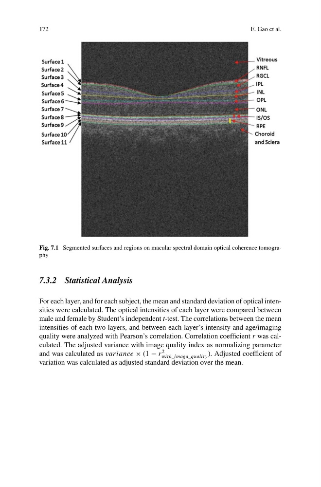
Fig. 7.1 Segmented surfaces and regions on macular spectral domain optical coherence tomography
7.3.2 Statistical Analysis
For each layer, and for each subject, the mean and standard deviation of optical intensities were calculated. The optical intensities of each layer were compared between
male and female by Student’s independent t-test. The correlations between the mean
intensities of each two layers, and between each layer’s intensity and age/imaging
quality were analyzed with Pearson’s correlation. Correlation coefficient r was calculated. The adjusted variance with image quality index as normalizing parameter
and was calculated as variance × (1 − r
2
with_imaga_qualit y ). Adjusted coefficient of
variation was calculated as adjusted standard deviation over the mean.
E. Gao et al.
Fig. 7.1 Segmented surfaces and regions on macular spectral domain optical coherence tomography
7.3.2 Statistical Analysis
For each layer, and for each subject, the mean and standard deviation of optical intensities were calculated. The optical intensities of each layer were compared between
male and female by Student’s independent t-test. The correlations between the mean
intensities of each two layers, and between each layer’s intensity and age/imaging
quality were analyzed with Pearson’s correlation. Correlation coefficient r was calculated. The adjusted variance with image quality index as normalizing parameter
and was calculated as variance × (1 − r
2
with_imaga_qualit y ). Adjusted coefficient of
variation was calculated as adjusted standard deviation over the mean.