

4
H. Chen et al.
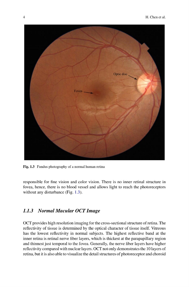
Fig. 1.3 Fundus photography of a normal human retina
responsible for fine vision and color vision. There is no inner retinal structure in
fovea, hence, there is no blood vessel and allows light to reach the photoreceptors
without any disturbance (Fig. 1.3).
1.1.3 Normal Macular OCT Image
OCT provides high resolution imaging for the cross-sectional structure of retina. The
reflectivity of tissue is determined by the optical character of tissue itself. Vitreous
has the lowest reflectivity in normal subjects. The highest reflective band at the
inner retina is retinal nerve fiber layers, which is thickest at the parapapillary region
and thinnest just temporal to the fovea. Generally, the nerve fiber layers have higher
reflectivity compared with nuclear layers. OCT not only demonstrates the 10 layers of
retina, but it is also able to visualize the detail structures of photoreceptor and choroid
H. Chen et al.
Fig. 1.3 Fundus photography of a normal human retina
responsible for fine vision and color vision. There is no inner retinal structure in
fovea, hence, there is no blood vessel and allows light to reach the photoreceptors
without any disturbance (Fig. 1.3).
1.1.3 Normal Macular OCT Image
OCT provides high resolution imaging for the cross-sectional structure of retina. The
reflectivity of tissue is determined by the optical character of tissue itself. Vitreous
has the lowest reflectivity in normal subjects. The highest reflective band at the
inner retina is retinal nerve fiber layers, which is thickest at the parapapillary region
and thinnest just temporal to the fovea. Generally, the nerve fiber layers have higher
reflectivity compared with nuclear layers. OCT not only demonstrates the 10 layers of
retina, but it is also able to visualize the detail structures of photoreceptor and choroid