

1 Clinical Applications of Retinal Optical Coherence Tomography
3
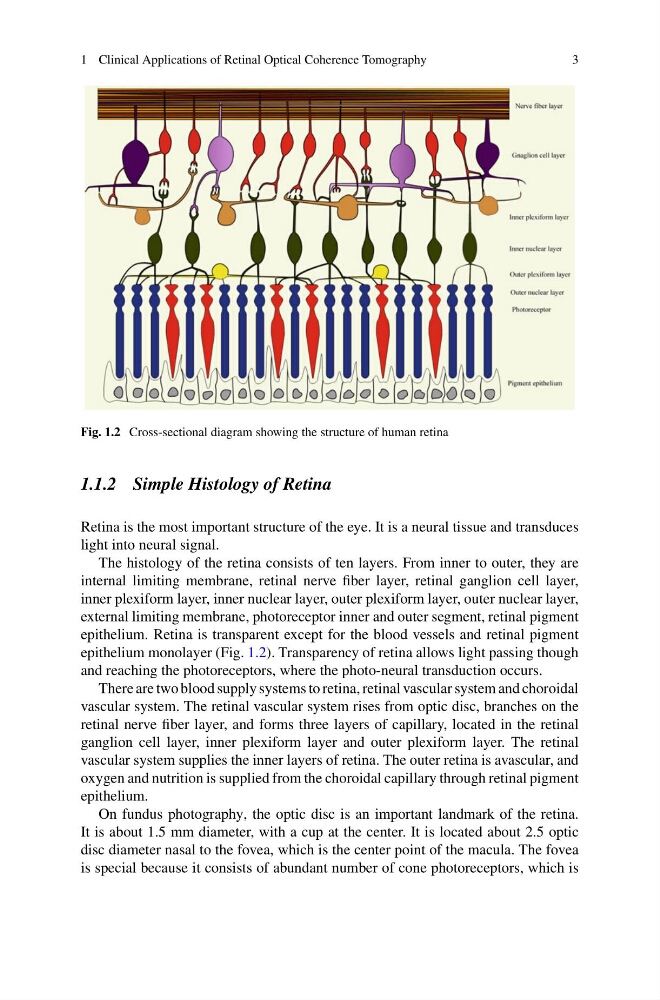
Fig. 1.2 Cross-sectional diagram showing the structure of human retina
1.1.2 Simple Histology of Retina
Retina is the most important structure of the eye. It is a neural tissue and transduces
light into neural signal.
The histology of the retina consists of ten layers. From inner to outer, they are
internal limiting membrane, retinal nerve fiber layer, retinal ganglion cell layer,
inner plexiform layer, inner nuclear layer, outer plexiform layer, outer nuclear layer,
external limiting membrane, photoreceptor inner and outer segment, retinal pigment
epithelium. Retina is transparent except for the blood vessels and retinal pigment
epithelium monolayer (Fig. 1.2). Transparency of retina allows light passing though
and reaching the photoreceptors, where the photo-neural transduction occurs.
There are two blood supply systems to retina, retinal vascular system and choroidal
vascular system. The retinal vascular system rises from optic disc, branches on the
retinal nerve fiber layer, and forms three layers of capillary, located in the retinal
ganglion cell layer, inner plexiform layer and outer plexiform layer. The retinal
vascular system supplies the inner layers of retina. The outer retina is avascular, and
oxygen and nutrition is supplied from the choroidal capillary through retinal pigment
epithelium.
On fundus photography, the optic disc is an important landmark of the retina.
It is about 1.5 mm diameter, with a cup at the center. It is located about 2.5 optic
disc diameter nasal to the fovea, which is the center point of the macula. The fovea
is special because it consists of abundant number of cone photoreceptors, which is
3
Fig. 1.2 Cross-sectional diagram showing the structure of human retina
1.1.2 Simple Histology of Retina
Retina is the most important structure of the eye. It is a neural tissue and transduces
light into neural signal.
The histology of the retina consists of ten layers. From inner to outer, they are
internal limiting membrane, retinal nerve fiber layer, retinal ganglion cell layer,
inner plexiform layer, inner nuclear layer, outer plexiform layer, outer nuclear layer,
external limiting membrane, photoreceptor inner and outer segment, retinal pigment
epithelium. Retina is transparent except for the blood vessels and retinal pigment
epithelium monolayer (Fig. 1.2). Transparency of retina allows light passing though
and reaching the photoreceptors, where the photo-neural transduction occurs.
There are two blood supply systems to retina, retinal vascular system and choroidal
vascular system. The retinal vascular system rises from optic disc, branches on the
retinal nerve fiber layer, and forms three layers of capillary, located in the retinal
ganglion cell layer, inner plexiform layer and outer plexiform layer. The retinal
vascular system supplies the inner layers of retina. The outer retina is avascular, and
oxygen and nutrition is supplied from the choroidal capillary through retinal pigment
epithelium.
On fundus photography, the optic disc is an important landmark of the retina.
It is about 1.5 mm diameter, with a cup at the center. It is located about 2.5 optic
disc diameter nasal to the fovea, which is the center point of the macula. The fovea
is special because it consists of abundant number of cone photoreceptors, which is