

354
epichlorohydrin due to the cross-linking reaction between the cyclodextrin molecules and epoxide and polymerized epichlorohydrin due to the homopolymerization
of epichlorohydrin with itself. These two components were analyzed in terms of
relaxation parameters, i.e.,
13
C spin lattice relaxation and
1
H spin lattice relaxation in
the rotating frame (Crini et al. 1998a, b, 2000).
In spite of the facile synthetic conditions for the preparation of ECP-based polymers, the polymer networks may adopt variable structural variability. Since cyclodextrin molecules contain several glucose units and hydroxyalkyl groups present at
positions 2, 3, and 6 in each glucose unit, the structure of the polymer network is
complicated because, during synthesis, many units can be interconnected as shown
in Fig. 8.2. This structure has been demonstrated by solid-state NMR experiments
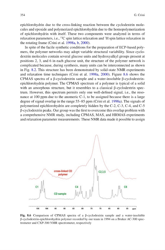
and relaxation time techniques (Crini et al. 1998a, 2000). Figure 8.6 shows the
CPMAS spectra of a β-cyclodextrin sample and a water-insoluble β-cyclodextrinepichlorohydrin polymer. The CPMAS spectrum of a polymer is typical of a solid
with an amorphous structure, but it resembles to a classical β-cyclodextrin spectrum. However, this spectrum permits only one well-defined signal, i.e., the resonance at 100 ppm due to the anomeric C-1, to be assigned because there is a large
degree of signal overlap in the range 55–85 ppm (Crini et al. 1998a). The signals of
polymerized epichlorohydrin are completely hidden by the C-2, C-3, C-4, and C-5
β-cyclodextrin peaks. Our group was the first to overcome this overlap problem with
a comprehensive NMR study, including CPMAS, MAS, and HRMAS experiments
and relaxation parameter measurements. These NMR data made it possible to assign
Fig. 8.6 Comparison of CPMAS spectra of a β-cyclodextrin sample and a water-insoluble
β-cyclodextrin-epichlorohydrin polymer recorded by our team in 1994 on a Bruker AC-300 spectrometer and CXP-300 NMR spectrometer, respectively
G. Crini
epichlorohydrin due to the cross-linking reaction between the cyclodextrin molecules and epoxide and polymerized epichlorohydrin due to the homopolymerization
of epichlorohydrin with itself. These two components were analyzed in terms of
relaxation parameters, i.e.,
13
C spin lattice relaxation and
1
H spin lattice relaxation in
the rotating frame (Crini et al. 1998a, b, 2000).
In spite of the facile synthetic conditions for the preparation of ECP-based polymers, the polymer networks may adopt variable structural variability. Since cyclodextrin molecules contain several glucose units and hydroxyalkyl groups present at
positions 2, 3, and 6 in each glucose unit, the structure of the polymer network is
complicated because, during synthesis, many units can be interconnected as shown
in Fig. 8.2. This structure has been demonstrated by solid-state NMR experiments
and relaxation time techniques (Crini et al. 1998a, 2000). Figure 8.6 shows the
CPMAS spectra of a β-cyclodextrin sample and a water-insoluble β-cyclodextrinepichlorohydrin polymer. The CPMAS spectrum of a polymer is typical of a solid
with an amorphous structure, but it resembles to a classical β-cyclodextrin spectrum. However, this spectrum permits only one well-defined signal, i.e., the resonance at 100 ppm due to the anomeric C-1, to be assigned because there is a large
degree of signal overlap in the range 55–85 ppm (Crini et al. 1998a). The signals of
polymerized epichlorohydrin are completely hidden by the C-2, C-3, C-4, and C-5
β-cyclodextrin peaks. Our group was the first to overcome this overlap problem with
a comprehensive NMR study, including CPMAS, MAS, and HRMAS experiments
and relaxation parameter measurements. These NMR data made it possible to assign
Fig. 8.6 Comparison of CPMAS spectra of a β-cyclodextrin sample and a water-insoluble
β-cyclodextrin-epichlorohydrin polymer recorded by our team in 1994 on a Bruker AC-300 spectrometer and CXP-300 NMR spectrometer, respectively
G. Crini