

1 3
Top Curr Chem (Z) (2018) 376:24
spectrum of the non-interacting dimer with the refined-mAS CAS(14,13)
δ
provides
a 2D map that is consistent with the sum of the 2D maps of the monomers computed at the CAS(6,6)
δ
and CAS(8,7)
δ
levels [58]. This preliminary check indicated
that the CAS(14,13)
δ
2DUV–UV spectrum of the non-interacting dimer, in turn,
could be used as a reference spectrum, to further reduce the computational cost.
The CAS(14,13)
δ
computation of the dimer, indeed, almost reaches the limit of a
reasonable computational time with current computer capabilities, and more efficient protocols would be beneficial. In this context, we have adopted the RASSCF
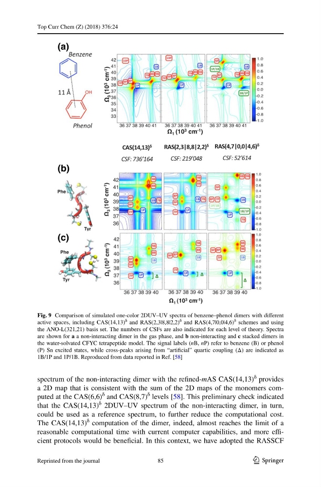
Fig. 9 Comparison of simulated one-color 2DUV–UV spectra of benzene–phenol dimers with different
active spaces, including CAS(14,13)
δ and RAS(2,3|8,8|2,2)
δ and RAS(4,7|0,0|4,6)
δ schemes and using
the ANO-L(321,21) basis set. The numbers of CSFs are also indicated for each level of theory. Spectra
are shown for a a non-interacting dimer in the gas phase, and b non-interacting and c stacked dimers in
the water-solvated CFYC tetrapeptide model. The signal labels (nB, nP) refer to benzene (B) or phenol
(P) Sn excited states, while cross-peaks arising from “artificial” quartic coupling (Δ) are indicated as
1B/1P and 1P/1B. Reproduced from data reported in Ref. [58]
85
Reprinted from the journal
Top Curr Chem (Z) (2018) 376:24
spectrum of the non-interacting dimer with the refined-mAS CAS(14,13)
δ
provides
a 2D map that is consistent with the sum of the 2D maps of the monomers computed at the CAS(6,6)
δ
and CAS(8,7)
δ
levels [58]. This preliminary check indicated
that the CAS(14,13)
δ
2DUV–UV spectrum of the non-interacting dimer, in turn,
could be used as a reference spectrum, to further reduce the computational cost.
The CAS(14,13)
δ
computation of the dimer, indeed, almost reaches the limit of a
reasonable computational time with current computer capabilities, and more efficient protocols would be beneficial. In this context, we have adopted the RASSCF
Fig. 9 Comparison of simulated one-color 2DUV–UV spectra of benzene–phenol dimers with different
active spaces, including CAS(14,13)
δ and RAS(2,3|8,8|2,2)
δ and RAS(4,7|0,0|4,6)
δ schemes and using
the ANO-L(321,21) basis set. The numbers of CSFs are also indicated for each level of theory. Spectra
are shown for a a non-interacting dimer in the gas phase, and b non-interacting and c stacked dimers in
the water-solvated CFYC tetrapeptide model. The signal labels (nB, nP) refer to benzene (B) or phenol
(P) Sn excited states, while cross-peaks arising from “artificial” quartic coupling (Δ) are indicated as
1B/1P and 1P/1B. Reproduced from data reported in Ref. [58]
85
Reprinted from the journal