

Top Curr Chem (Z) (2018) 376:10
1 3
gives origin to vibronic (excited states) and vibrational (ground state) coherences,
which can both lead to long-lived beating signals in 2DES maps.
By retrieving the 2D Fourier-transform amplitude maps of selected oscillation at
frequency ν 1 close to 700 cm
−1
, the authors observed a very defined coherence pattern for the non-rephasing and rephasing spectra. Specifically, the oscillations appear
on the diagonal peak for the non-rephasing map and as a cross-peak on the rephasing map (Fig. 14c). To describe these features, a vibronic model is employed that
describes the coupling of the two electronic bands (specifically 1 and 3 in Fig. 14b)
with a quasi-resonant vibrational mode with frequency ν 1 . The model predicts an
initial fast decoherence, sustained by a long-lived vibrational character of the underdamped vibration (Fig. 14d). The polarization-implemented sequence of this 2DES
result provides a strong foundation for understating vibronic coupling and its implication in energy transfer processes.
Coherent oscillations are clearly observed and assigned in inorganic complexes
as well, which are characterized by narrower spectral lineshapes and reduction of
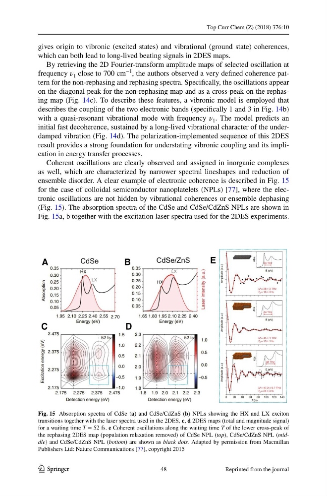
ensemble disorder. A clear example of electronic coherence is described in Fig. 15
for the case of colloidal semiconductor nanoplatelets (NPLs) [77], where the electronic oscillations are not hidden by vibrational coherences or ensemble dephasing
(Fig. 15). The absorption spectra of the CdSe and CdSe/CdZnS NPLs are shown in
Fig. 15a, b together with the excitation laser spectra used for the 2DES experiments.
Fig. 15 Absorption spectra of CdSe (a) and CdSe/CdZnS (b) NPLs showing the HX and LX exciton
transitions together with the laser spectra used in the 2DES. c, d 2DES maps (total and magnitude signal)
for a waiting time T = 52 fs. e Coherent oscillations along the waiting time T of the lower cross-peak of
the rephasing 2DES map (population relaxation removed) of CdSe NPL (top), CdSe/CdZnS NPL (middle) and CdSe/CdZnS NPL (bottom) are shown as black dots. Adapted by permission from Macmillan
Publishers Ltd: Nature Communications [77], copyright 2015
48
Reprinted from the journal
1 3
gives origin to vibronic (excited states) and vibrational (ground state) coherences,
which can both lead to long-lived beating signals in 2DES maps.
By retrieving the 2D Fourier-transform amplitude maps of selected oscillation at
frequency ν 1 close to 700 cm
−1
, the authors observed a very defined coherence pattern for the non-rephasing and rephasing spectra. Specifically, the oscillations appear
on the diagonal peak for the non-rephasing map and as a cross-peak on the rephasing map (Fig. 14c). To describe these features, a vibronic model is employed that
describes the coupling of the two electronic bands (specifically 1 and 3 in Fig. 14b)
with a quasi-resonant vibrational mode with frequency ν 1 . The model predicts an
initial fast decoherence, sustained by a long-lived vibrational character of the underdamped vibration (Fig. 14d). The polarization-implemented sequence of this 2DES
result provides a strong foundation for understating vibronic coupling and its implication in energy transfer processes.
Coherent oscillations are clearly observed and assigned in inorganic complexes
as well, which are characterized by narrower spectral lineshapes and reduction of
ensemble disorder. A clear example of electronic coherence is described in Fig. 15
for the case of colloidal semiconductor nanoplatelets (NPLs) [77], where the electronic oscillations are not hidden by vibrational coherences or ensemble dephasing
(Fig. 15). The absorption spectra of the CdSe and CdSe/CdZnS NPLs are shown in
Fig. 15a, b together with the excitation laser spectra used for the 2DES experiments.
Fig. 15 Absorption spectra of CdSe (a) and CdSe/CdZnS (b) NPLs showing the HX and LX exciton
transitions together with the laser spectra used in the 2DES. c, d 2DES maps (total and magnitude signal)
for a waiting time T = 52 fs. e Coherent oscillations along the waiting time T of the lower cross-peak of
the rephasing 2DES map (population relaxation removed) of CdSe NPL (top), CdSe/CdZnS NPL (middle) and CdSe/CdZnS NPL (bottom) are shown as black dots. Adapted by permission from Macmillan
Publishers Ltd: Nature Communications [77], copyright 2015
48
Reprinted from the journal