

1 3
Top Curr Chem (Z) (2018) 376:10
components. In this system, a quasi-resonance exists between the vibrational frequencies ν 1 and ν 2 and the energy splitting of the electronic transition, which, in
principle, makes it complicated to distinguish the contribution from electronic or
vibrational origin. The resonance promotes an electronic–vibrational coupling that
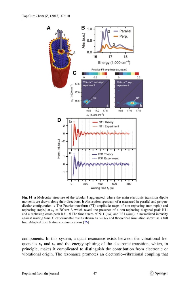
Fig. 14 a Molecular structure of the tubular J aggregated, where the main electronic transition dipole
moments are drawn along their directions. b Absorption spectrum of a measured in parallel and perpendicular configuration. c The Fourier-transform (FT) amplitude maps of non-rephasing (non-reph.) and
rephasing (reph.) at ν 1 = 700 cm
−1 , which reveal the presence of a non-rephasing diagonal peak N11
and a rephasing cross-peak R31. d The time traces of N11 (red) and R31 (blue) in normalized intensity
against waiting time T: experimental results shown as circles and theoretical simulation shown as a full
line. Adapted from Nature communications [76]
47
Reprinted from the journal
Top Curr Chem (Z) (2018) 376:10
components. In this system, a quasi-resonance exists between the vibrational frequencies ν 1 and ν 2 and the energy splitting of the electronic transition, which, in
principle, makes it complicated to distinguish the contribution from electronic or
vibrational origin. The resonance promotes an electronic–vibrational coupling that
Fig. 14 a Molecular structure of the tubular J aggregated, where the main electronic transition dipole
moments are drawn along their directions. b Absorption spectrum of a measured in parallel and perpendicular configuration. c The Fourier-transform (FT) amplitude maps of non-rephasing (non-reph.) and
rephasing (reph.) at ν 1 = 700 cm
−1 , which reveal the presence of a non-rephasing diagonal peak N11
and a rephasing cross-peak R31. d The time traces of N11 (red) and R31 (blue) in normalized intensity
against waiting time T: experimental results shown as circles and theoretical simulation shown as a full
line. Adapted from Nature communications [76]
47
Reprinted from the journal