

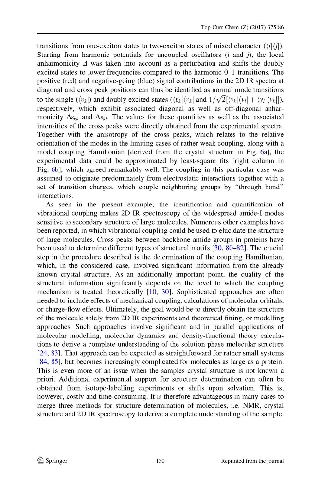
transitions from one-exciton states to two-exciton states of mixed character (hijhjj).
Starting from harmonic potentials for uncoupled oscillators (i and j), the local
anharmonicity D was taken into account as a perturbation and shifts the doubly
excited states to lower frequencies compared to the harmonic 0–1 transitions. The
positive (red) and negative-going (blue) signal contributions in the 2D IR spectra at
diagonal and cross peak positions can thus be identified as normal mode transitions
to the single (ht k j) and doubly excited states (ht k jht k j and 1=
ffiffi ffi
2
p
hm k jhm l j þ hm l jhm k j
½
),
respectively, which exhibit associated diagonal as well as off-diagonal anharmonicity De kk and De kl . The values for these quantities as well as the associated
intensities of the cross peaks were directly obtained from the experimental spectra.
Together with the anisotropy of the cross peaks, which relates to the relative
orientation of the modes in the limiting cases of rather weak coupling, along with a
model coupling Hamiltonian [derived from the crystal structure in Fig. 6a], the
experimental data could be approximated by least-square fits [right column in
Fig. 6b], which agreed remarkably well. The coupling in this particular case was
assumed to originate predominately from electrostatic interactions together with a
set of transition charges, which couple neighboring groups by ‘‘through bond’’
interactions.
As seen in the present example, the identification and quantification of
vibrational coupling makes 2D IR spectroscopy of the widespread amide-I modes
sensitive to secondary structure of large molecules. Numerous other examples have
been reported, in which vibrational coupling could be used to elucidate the structure
of large molecules. Cross peaks between backbone amide groups in proteins have
been used to determine different types of structural motifs [30, 80–82]. The crucial
step in the procedure described is the determination of the coupling Hamiltonian,
which, in the considered case, involved significant information from the already
known crystal structure. As an additionally important point, the quality of the
structural information significantly depends on the level to which the coupling
mechanism is treated theoretically [10, 30]. Sophisticated approaches are often
needed to include effects of mechanical coupling, calculations of molecular orbitals,
or charge-flow effects. Ultimately, the goal would be to directly obtain the structure
of the molecule solely from 2D IR experiments and theoretical fitting, or modelling
approaches. Such approaches involve significant and in parallel applications of
molecular modelling, molecular dynamics and density-functional theory calculations to derive a complete understanding of the solution phase molecular structure
[24, 83]. That approach can be expected as straightforward for rather small systems
[84, 85], but becomes increasingly complicated for molecules as large as a protein.
This is even more of an issue when the samples crystal structure is not known a
priori. Additional experimental support for structure determination can often be
obtained from isotope-labelling experiments or shifts upon solvation. This is,
however, costly and time-consuming. It is therefore advantageous in many cases to
merge three methods for structure determination of molecules, i.e. NMR, crystal
structure and 2D IR spectroscopy to derive a complete understanding of the sample.
Top Curr Chem (Z) (2017) 375:86
123
130
Reprinted from the journal
Starting from harmonic potentials for uncoupled oscillators (i and j), the local
anharmonicity D was taken into account as a perturbation and shifts the doubly
excited states to lower frequencies compared to the harmonic 0–1 transitions. The
positive (red) and negative-going (blue) signal contributions in the 2D IR spectra at
diagonal and cross peak positions can thus be identified as normal mode transitions
to the single (ht k j) and doubly excited states (ht k jht k j and 1=
ffiffi ffi
2
p
hm k jhm l j þ hm l jhm k j
½
),
respectively, which exhibit associated diagonal as well as off-diagonal anharmonicity De kk and De kl . The values for these quantities as well as the associated
intensities of the cross peaks were directly obtained from the experimental spectra.
Together with the anisotropy of the cross peaks, which relates to the relative
orientation of the modes in the limiting cases of rather weak coupling, along with a
model coupling Hamiltonian [derived from the crystal structure in Fig. 6a], the
experimental data could be approximated by least-square fits [right column in
Fig. 6b], which agreed remarkably well. The coupling in this particular case was
assumed to originate predominately from electrostatic interactions together with a
set of transition charges, which couple neighboring groups by ‘‘through bond’’
interactions.
As seen in the present example, the identification and quantification of
vibrational coupling makes 2D IR spectroscopy of the widespread amide-I modes
sensitive to secondary structure of large molecules. Numerous other examples have
been reported, in which vibrational coupling could be used to elucidate the structure
of large molecules. Cross peaks between backbone amide groups in proteins have
been used to determine different types of structural motifs [30, 80–82]. The crucial
step in the procedure described is the determination of the coupling Hamiltonian,
which, in the considered case, involved significant information from the already
known crystal structure. As an additionally important point, the quality of the
structural information significantly depends on the level to which the coupling
mechanism is treated theoretically [10, 30]. Sophisticated approaches are often
needed to include effects of mechanical coupling, calculations of molecular orbitals,
or charge-flow effects. Ultimately, the goal would be to directly obtain the structure
of the molecule solely from 2D IR experiments and theoretical fitting, or modelling
approaches. Such approaches involve significant and in parallel applications of
molecular modelling, molecular dynamics and density-functional theory calculations to derive a complete understanding of the solution phase molecular structure
[24, 83]. That approach can be expected as straightforward for rather small systems
[84, 85], but becomes increasingly complicated for molecules as large as a protein.
This is even more of an issue when the samples crystal structure is not known a
priori. Additional experimental support for structure determination can often be
obtained from isotope-labelling experiments or shifts upon solvation. This is,
however, costly and time-consuming. It is therefore advantageous in many cases to
merge three methods for structure determination of molecules, i.e. NMR, crystal
structure and 2D IR spectroscopy to derive a complete understanding of the sample.
Top Curr Chem (Z) (2017) 375:86
123
130
Reprinted from the journal