

ultrashort laser pulses, which initially excite a molecular vibration and successively
interrogate the evolution of the initial excitation by the help of a time-delayed probe
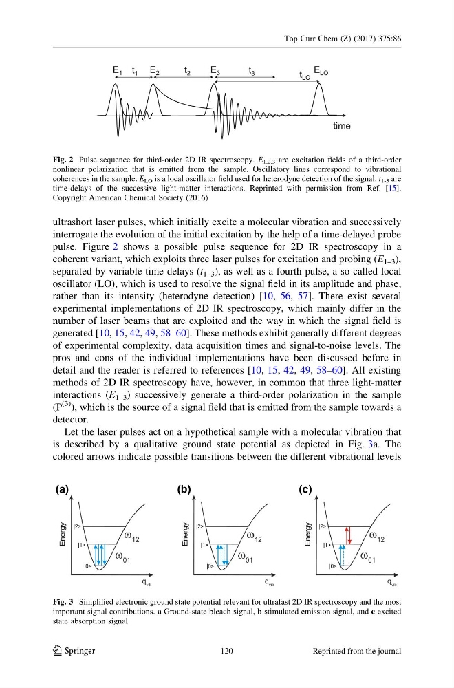
pulse. Figure 2 shows a possible pulse sequence for 2D IR spectroscopy in a
coherent variant, which exploits three laser pulses for excitation and probing (E 1–3 ),
separated by variable time delays (t 1–3 ), as well as a fourth pulse, a so-called local
oscillator (LO), which is used to resolve the signal field in its amplitude and phase,
rather than its intensity (heterodyne detection) [10, 56, 57]. There exist several
experimental implementations of 2D IR spectroscopy, which mainly differ in the
number of laser beams that are exploited and the way in which the signal field is
generated [10, 15, 42, 49, 58–60]. These methods exhibit generally different degrees
of experimental complexity, data acquisition times and signal-to-noise levels. The
pros and cons of the individual implementations have been discussed before in
detail and the reader is referred to references [10, 15, 42, 49, 58–60]. All existing
methods of 2D IR spectroscopy have, however, in common that three light-matter
interactions (E 1–3 ) successively generate a third-order polarization in the sample
(P
(3) ), which is the source of a signal field that is emitted from the sample towards a
detector.
Let the laser pulses act on a hypothetical sample with a molecular vibration that
is described by a qualitative ground state potential as depicted in Fig. 3a. The
colored arrows indicate possible transitions between the different vibrational levels
E 1
E 2
E 3
time
E LO
t 1
t 2
t 3
t LO
Fig. 2 Pulse sequence for third-order 2D IR spectroscopy. E 1,2,3 are excitation fields of a third-order
nonlinear polarization that is emitted from the sample. Oscillatory lines correspond to vibrational
coherences in the sample. E LO is a local oscillator field used for heterodyne detection of the signal. t 1-3 are
time-delays of the successive light-matter interactions. Reprinted with permission from Ref. [15].
Copyright American Chemical Society (2016)
(a)
(b)
(c)
Fig. 3 Simplified electronic ground state potential relevant for ultrafast 2D IR spectroscopy and the most
important signal contributions. a Ground-state bleach signal, b stimulated emission signal, and c excited
state absorption signal
Top Curr Chem (Z) (2017) 375:86
123
120
Reprinted from the journal
interrogate the evolution of the initial excitation by the help of a time-delayed probe
pulse. Figure 2 shows a possible pulse sequence for 2D IR spectroscopy in a
coherent variant, which exploits three laser pulses for excitation and probing (E 1–3 ),
separated by variable time delays (t 1–3 ), as well as a fourth pulse, a so-called local
oscillator (LO), which is used to resolve the signal field in its amplitude and phase,
rather than its intensity (heterodyne detection) [10, 56, 57]. There exist several
experimental implementations of 2D IR spectroscopy, which mainly differ in the
number of laser beams that are exploited and the way in which the signal field is
generated [10, 15, 42, 49, 58–60]. These methods exhibit generally different degrees
of experimental complexity, data acquisition times and signal-to-noise levels. The
pros and cons of the individual implementations have been discussed before in
detail and the reader is referred to references [10, 15, 42, 49, 58–60]. All existing
methods of 2D IR spectroscopy have, however, in common that three light-matter
interactions (E 1–3 ) successively generate a third-order polarization in the sample
(P
(3) ), which is the source of a signal field that is emitted from the sample towards a
detector.
Let the laser pulses act on a hypothetical sample with a molecular vibration that
is described by a qualitative ground state potential as depicted in Fig. 3a. The
colored arrows indicate possible transitions between the different vibrational levels
E 1
E 2
E 3
time
E LO
t 1
t 2
t 3
t LO
Fig. 2 Pulse sequence for third-order 2D IR spectroscopy. E 1,2,3 are excitation fields of a third-order
nonlinear polarization that is emitted from the sample. Oscillatory lines correspond to vibrational
coherences in the sample. E LO is a local oscillator field used for heterodyne detection of the signal. t 1-3 are
time-delays of the successive light-matter interactions. Reprinted with permission from Ref. [15].
Copyright American Chemical Society (2016)
(a)
(b)
(c)
Fig. 3 Simplified electronic ground state potential relevant for ultrafast 2D IR spectroscopy and the most
important signal contributions. a Ground-state bleach signal, b stimulated emission signal, and c excited
state absorption signal
Top Curr Chem (Z) (2017) 375:86
123
120
Reprinted from the journal