

These include the straightforward incorporation of chemical functionality by use of
functional amino acids, as well as enzymatic degradability.
Following up on this work, Sofroniew, Deming and colleagues studied the
biocompatibility of diblock copolypeptide hydrogels in vivo in mouse central
nervous system (CNS) tissue [121]. This work was undertaken because biomaterials represent a major opportunity for developing novel CNS treatment strategies
based on site-specific delivery of scaffolds that promote the growth and migration
of axons or cells derived from host or grafts, or as depots that release diffusible
bioactive molecules to act in a locally restricted manner inside the blood–brain
barrier. A range of diblock copolypeptide hydrogel formulations with rheological
properties similar to brain tissue were injected into mouse forebrain and examined
after 1–8 weeks using light microscopy, immunohistochemistry, and electron
microscopy. Hydrogel deposits were found to elicit no more gliosis, inflammation,
or toxicity to neurons, myelin, or axons than did injections of physiological saline.
The size, rigidity, and density of the hydrogel deposits could be varied subtly by
altering sample composition and concentration. The K 180 L 20 hydrogel was selected
for detailed analyses because it formed deposits with desirable physical properties
and because lysine is routinely used as a substrate for neural cell cultures. Deposits
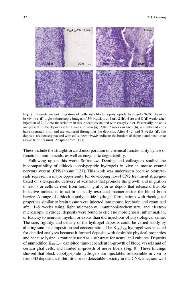
of unmodified K 180 L 20 exhibited time-dependent in-growth of blood vessels and of
certain glial cells, and limited in-growth of nerve fibers (Fig. 8). These findings
showed that block copolypeptide hydrogels are injectable, re-assemble in vivo to
form 3D deposits, exhibit little or no detectable toxicity in the CNS, integrate well
Fig. 8 Time-dependent migration of cells into block copolypeptide hydrogel (DCH) deposits
in vivo. (a–d) Light-microscopic images of 3% K 180 L 20 at 1 (a), 2 (b), 4 (c) and 8 (d) weeks after
injection of 2 μL into the striatum in tissue sections stained with cresyl violet. Essentially, no cells
are present in the deposits after 1 week in vivo (a). After 2 weeks in vivo (b), a number of cells
have migrated into, and are scattered throughout the deposits. After 4 (c) and 8 weeks (d), the
deposits are densely packed with cells. Arrowheads indicate the borders of deposit and host tissue
(scale bars: 25 mm). Adapted from [121]
32
T.J. Deming
functional amino acids, as well as enzymatic degradability.
Following up on this work, Sofroniew, Deming and colleagues studied the
biocompatibility of diblock copolypeptide hydrogels in vivo in mouse central
nervous system (CNS) tissue [121]. This work was undertaken because biomaterials represent a major opportunity for developing novel CNS treatment strategies
based on site-specific delivery of scaffolds that promote the growth and migration
of axons or cells derived from host or grafts, or as depots that release diffusible
bioactive molecules to act in a locally restricted manner inside the blood–brain
barrier. A range of diblock copolypeptide hydrogel formulations with rheological
properties similar to brain tissue were injected into mouse forebrain and examined
after 1–8 weeks using light microscopy, immunohistochemistry, and electron
microscopy. Hydrogel deposits were found to elicit no more gliosis, inflammation,
or toxicity to neurons, myelin, or axons than did injections of physiological saline.
The size, rigidity, and density of the hydrogel deposits could be varied subtly by
altering sample composition and concentration. The K 180 L 20 hydrogel was selected
for detailed analyses because it formed deposits with desirable physical properties
and because lysine is routinely used as a substrate for neural cell cultures. Deposits
of unmodified K 180 L 20 exhibited time-dependent in-growth of blood vessels and of
certain glial cells, and limited in-growth of nerve fibers (Fig. 8). These findings
showed that block copolypeptide hydrogels are injectable, re-assemble in vivo to
form 3D deposits, exhibit little or no detectable toxicity in the CNS, integrate well
Fig. 8 Time-dependent migration of cells into block copolypeptide hydrogel (DCH) deposits
in vivo. (a–d) Light-microscopic images of 3% K 180 L 20 at 1 (a), 2 (b), 4 (c) and 8 (d) weeks after
injection of 2 μL into the striatum in tissue sections stained with cresyl violet. Essentially, no cells
are present in the deposits after 1 week in vivo (a). After 2 weeks in vivo (b), a number of cells
have migrated into, and are scattered throughout the deposits. After 4 (c) and 8 weeks (d), the
deposits are densely packed with cells. Arrowheads indicate the borders of deposit and host tissue
(scale bars: 25 mm). Adapted from [121]
32
T.J. Deming