

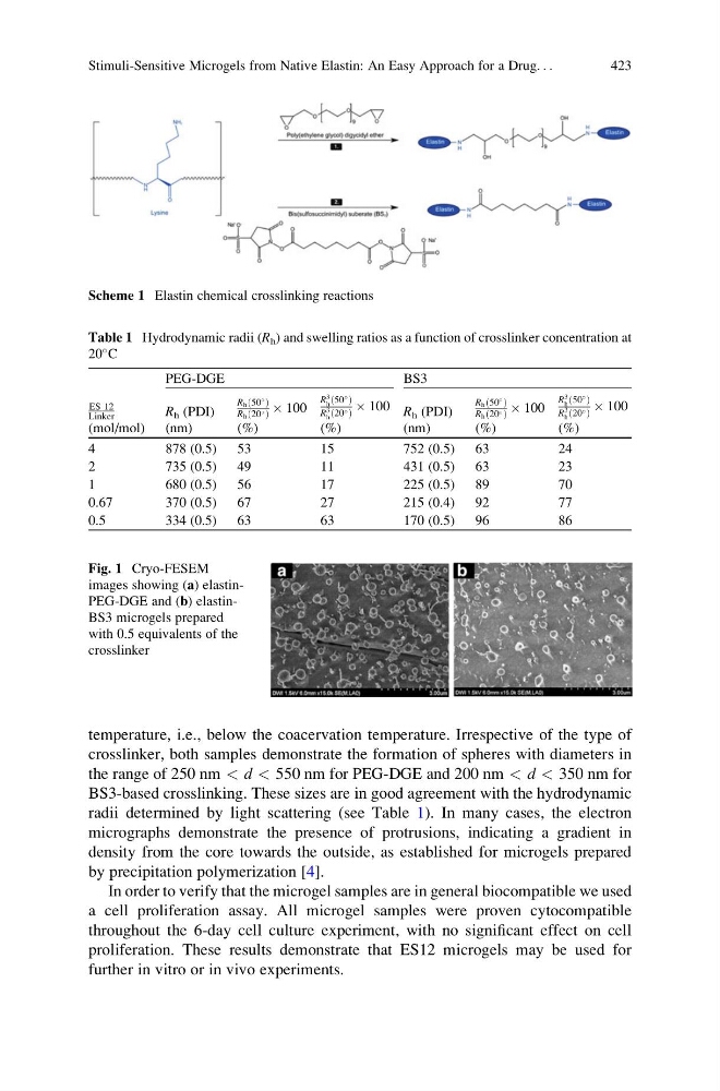
temperature, i.e., below the coacervation temperature. Irrespective of the type of
crosslinker, both samples demonstrate the formation of spheres with diameters in
the range of 250 nm < d < 550 nm for PEG-DGE and 200 nm < d < 350 nm for
BS3-based crosslinking. These sizes are in good agreement with the hydrodynamic
radii determined by light scattering (see Table 1). In many cases, the electron
micrographs demonstrate the presence of protrusions, indicating a gradient in
density from the core towards the outside, as established for microgels prepared
by precipitation polymerization [4].
In order to verify that the microgel samples are in general biocompatible we used
a cell proliferation assay. All microgel samples were proven cytocompatible
throughout the 6-day cell culture experiment, with no significant effect on cell
proliferation. These results demonstrate that ES12 microgels may be used for
further in vitro or in vivo experiments.
Table 1 Hydrodynamic radii (R h ) and swelling ratios as a function of crosslinker concentration at
20
C
ES 12
Linker
(mol/mol)
PEG-DGE
BS3
R h (PDI)
(nm)
Rh 50
ð Þ
Rh 20
ð Þ Â 100
(%)
R
3
h 50
ð Þ
R
3
h 20
ð Þ
 100
(%)
R h (PDI)
(nm)
Rh 50
ð Þ
Rh 20
ð Þ Â 100
(%)
R
3
h 50
ð Þ
R
3
h 20
ð Þ
 100
(%)
4
878 (0.5) 53
15
752 (0.5) 63
24
2
735 (0.5) 49
11
431 (0.5) 63
23
1
680 (0.5) 56
17
225 (0.5) 89
70
0.67
370 (0.5) 67
27
215 (0.4) 92
77
0.5
334 (0.5) 63
63
170 (0.5) 96
86
Fig. 1 Cryo-FESEM
images showing (a) elastinPEG-DGE and (b) elastinBS3 microgels prepared
with 0.5 equivalents of the
crosslinker
Scheme 1 Elastin chemical crosslinking reactions
Stimuli-Sensitive Microgels from Native Elastin: An Easy Approach for a Drug. . .
423
crosslinker, both samples demonstrate the formation of spheres with diameters in
the range of 250 nm < d < 550 nm for PEG-DGE and 200 nm < d < 350 nm for
BS3-based crosslinking. These sizes are in good agreement with the hydrodynamic
radii determined by light scattering (see Table 1). In many cases, the electron
micrographs demonstrate the presence of protrusions, indicating a gradient in
density from the core towards the outside, as established for microgels prepared
by precipitation polymerization [4].
In order to verify that the microgel samples are in general biocompatible we used
a cell proliferation assay. All microgel samples were proven cytocompatible
throughout the 6-day cell culture experiment, with no significant effect on cell
proliferation. These results demonstrate that ES12 microgels may be used for
further in vitro or in vivo experiments.
Table 1 Hydrodynamic radii (R h ) and swelling ratios as a function of crosslinker concentration at
20
C
ES 12
Linker
(mol/mol)
PEG-DGE
BS3
R h (PDI)
(nm)
Rh 50
ð Þ
Rh 20
ð Þ Â 100
(%)
R
3
h 50
ð Þ
R
3
h 20
ð Þ
 100
(%)
R h (PDI)
(nm)
Rh 50
ð Þ
Rh 20
ð Þ Â 100
(%)
R
3
h 50
ð Þ
R
3
h 20
ð Þ
 100
(%)
4
878 (0.5) 53
15
752 (0.5) 63
24
2
735 (0.5) 49
11
431 (0.5) 63
23
1
680 (0.5) 56
17
225 (0.5) 89
70
0.67
370 (0.5) 67
27
215 (0.4) 92
77
0.5
334 (0.5) 63
63
170 (0.5) 96
86
Fig. 1 Cryo-FESEM
images showing (a) elastinPEG-DGE and (b) elastinBS3 microgels prepared
with 0.5 equivalents of the
crosslinker
Scheme 1 Elastin chemical crosslinking reactions
Stimuli-Sensitive Microgels from Native Elastin: An Easy Approach for a Drug. . .
423