

show an increase in the observed fluorescence intensity. Thus, only about 3% of the
prepared peptoids were considered a hit. Size-exclusion chromatography confirmed
that these structures formed aggregates with aggregation numbers of 3–4 whereas
control peptoids did not.
This concept was later refined when several helices were covalently linked. It
was found, using Fo ¨rster resonance energy transfer (FRET), that these peptoids
undergo a cooperative transition into aggregates with a hydrophobic core that was
tentatively assigned to a tertiary structure similar to that found in folded proteins
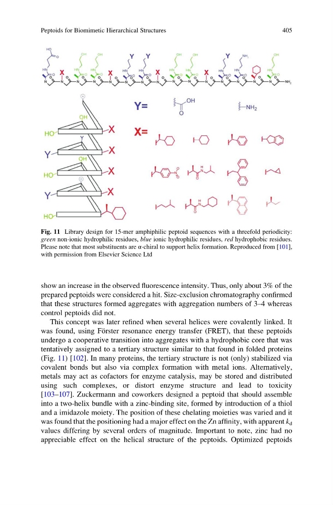
(Fig. 11) [102]. In many proteins, the tertiary structure is not (only) stabilized via
covalent bonds but also via complex formation with metal ions. Alternatively,
metals may act as cofactors for enzyme catalysis, may be stored and distributed
using such complexes, or distort enzyme structure and lead to toxicity
[103–107]. Zuckermann and coworkers designed a peptoid that should assemble
into a two-helix bundle with a zinc-binding site, formed by introduction of a thiol
and a imidazole moiety. The position of these chelating moieties was varied and it
was found that the positioning had a major effect on the Zn affinity, with apparent k d
values differing by several orders of magnitude. Important to note, zinc had no
appreciable effect on the helical structure of the peptoids. Optimized peptoids
Fig. 11 Library design for 15-mer amphiphilic peptoid sequences with a threefold periodicity:
green non-ionic hydrophilic residues, blue ionic hydrophilic residues, red hydrophobic residues.
Please note that most substituents are α-chiral to support helix formation. Reproduced from [101],
with permission from Elsevier Science Ltd
Peptoids for Biomimetic Hierarchical Structures
405
prepared peptoids were considered a hit. Size-exclusion chromatography confirmed
that these structures formed aggregates with aggregation numbers of 3–4 whereas
control peptoids did not.
This concept was later refined when several helices were covalently linked. It
was found, using Fo ¨rster resonance energy transfer (FRET), that these peptoids
undergo a cooperative transition into aggregates with a hydrophobic core that was
tentatively assigned to a tertiary structure similar to that found in folded proteins
(Fig. 11) [102]. In many proteins, the tertiary structure is not (only) stabilized via
covalent bonds but also via complex formation with metal ions. Alternatively,
metals may act as cofactors for enzyme catalysis, may be stored and distributed
using such complexes, or distort enzyme structure and lead to toxicity
[103–107]. Zuckermann and coworkers designed a peptoid that should assemble
into a two-helix bundle with a zinc-binding site, formed by introduction of a thiol
and a imidazole moiety. The position of these chelating moieties was varied and it
was found that the positioning had a major effect on the Zn affinity, with apparent k d
values differing by several orders of magnitude. Important to note, zinc had no
appreciable effect on the helical structure of the peptoids. Optimized peptoids
Fig. 11 Library design for 15-mer amphiphilic peptoid sequences with a threefold periodicity:
green non-ionic hydrophilic residues, blue ionic hydrophilic residues, red hydrophobic residues.
Please note that most substituents are α-chiral to support helix formation. Reproduced from [101],
with permission from Elsevier Science Ltd
Peptoids for Biomimetic Hierarchical Structures
405