

helices are observed for oligomers as short as five monomer units and the stability
increases up to DP ¼ 15. Interestingly, about 50% of the monomer units must carry
α-chiral side chains for helices to form [60–62]. This value in fact fits very well with
observations made in 1963 by Fasman and Blout for copolymers of prolin
(helicogenic) and sarcosine (non-helicogenic) [63].
Secondary structures such as helices are not restricted to uniform peptoids but
have also been described for non-uniform peptoids and structural isomers
[47, 64]. Similar to the early reports on helical peptoids, this was achieved by
introducing chiral side chains. Interestingly, linear peptoids yielded less intense
circular dichroism as compared to cyclic ones, suggesting less stable helices.
Peptoid helices are not stabilized by H-bonding. Regarding the stability of
peptoid helices, a mixed impression can be gained from the literature. On the one
hand, peptoid helices are very stable against typical denaturants, such as urea and
temperature [65]. On the other hand, conformational stability is typically less than
that of α-helices. This is evidenced by a rather low persistence length of peptoid
helices and low K cis/trans values [60]. Spectroscopic evidence also shows that polyL-proline helices are less rigid in solution than previously suggested [66, 67]. Similarly, the persistence length of the helices from C α -chiral peptoids were found to
range between 4 and 1 nm, depending on the chain length. Larger persistence
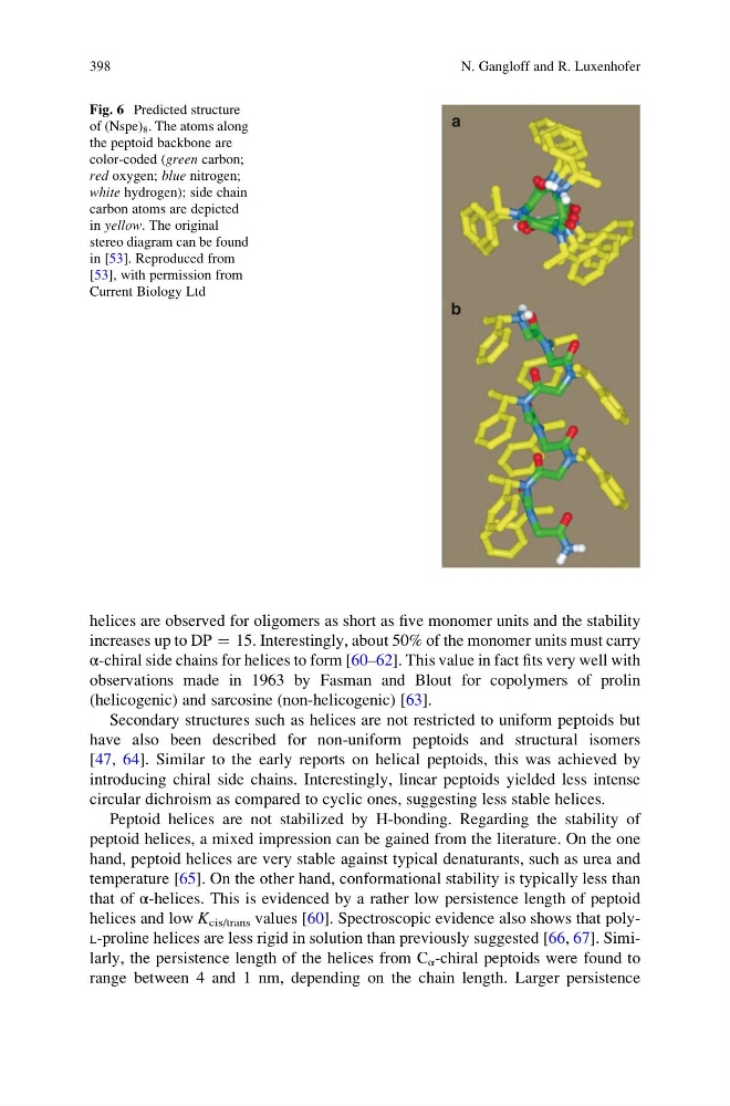
Fig. 6 Predicted structure
of (Nspe) 8 . The atoms along
the peptoid backbone are
color-coded (green carbon;
red oxygen; blue nitrogen;
white hydrogen); side chain
carbon atoms are depicted
in yellow. The original
stereo diagram can be found
in [53]. Reproduced from
[53], with permission from
Current Biology Ltd
398
N. Gangloff and R. Luxenhofer
increases up to DP ¼ 15. Interestingly, about 50% of the monomer units must carry
α-chiral side chains for helices to form [60–62]. This value in fact fits very well with
observations made in 1963 by Fasman and Blout for copolymers of prolin
(helicogenic) and sarcosine (non-helicogenic) [63].
Secondary structures such as helices are not restricted to uniform peptoids but
have also been described for non-uniform peptoids and structural isomers
[47, 64]. Similar to the early reports on helical peptoids, this was achieved by
introducing chiral side chains. Interestingly, linear peptoids yielded less intense
circular dichroism as compared to cyclic ones, suggesting less stable helices.
Peptoid helices are not stabilized by H-bonding. Regarding the stability of
peptoid helices, a mixed impression can be gained from the literature. On the one
hand, peptoid helices are very stable against typical denaturants, such as urea and
temperature [65]. On the other hand, conformational stability is typically less than
that of α-helices. This is evidenced by a rather low persistence length of peptoid
helices and low K cis/trans values [60]. Spectroscopic evidence also shows that polyL-proline helices are less rigid in solution than previously suggested [66, 67]. Similarly, the persistence length of the helices from C α -chiral peptoids were found to
range between 4 and 1 nm, depending on the chain length. Larger persistence
Fig. 6 Predicted structure
of (Nspe) 8 . The atoms along
the peptoid backbone are
color-coded (green carbon;
red oxygen; blue nitrogen;
white hydrogen); side chain
carbon atoms are depicted
in yellow. The original
stereo diagram can be found
in [53]. Reproduced from
[53], with permission from
Current Biology Ltd
398
N. Gangloff and R. Luxenhofer