

through a ~ 1 eV resolution cryogenically cooled diamond or Si pre-monochromator
(Chap. 4). A high-resolution monochromator using asymmetrically cut crystals then
narrows the bandpass to ~1 meV. Finally, the beam impinges on a sample that is
usually maintained at low temperature in a liquid nitrogen or liquid He cryostat.
The APD detector usually has a time resolution on the order ~1 ns, but it takes
multiple time constants to relax to baseline after the strong prompt pulse. A time gate
is set on the output from the APD, so that counts are only recorded after a
predetermined delay with respect to the prompt radiation (Fig. 10.3). In this manner,
the NRVS signal, consisting of delayed nuclear fluorescence and X-rays
(or electrons) following relaxation by internal conversion, is separated from purely
electronic events such as Rayleigh and Compton scattering and normal X-ray
fluorescence.
Not every Mössbauer isotope is suitable for an NRVS experiment. First of all, one
needs a monochromator with sufficiently narrow bandpass to resolve vibrational
features, generally ~meV resolution. At the moment, this limits the nuclear resonance energy to <40 keV. Second, the nuclear excited-state lifetime has to be long
enough (a few hundred ps) to allow the detector to distinguish nuclear events from
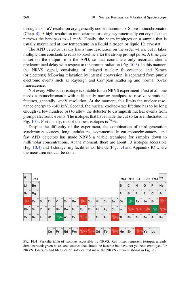
prompt electronic events. The isotopes that have made the cut so far are illustrated in
Fig. 10.4. Fortunately, one of the best isotopes is
57 Fe.
Despite the difficulty of the experiment, the combination of third-generation
synchrotron sources, long undulators, asymmetrically cut monochromators, and
fast APD detectors has made NRVS a viable technique for samples down to
millimolar concentrations. At the moment, there are about 13 isotopes accessible
(Fig. 10.4) and 4 storage ring facilities worldwide (Fig. 1.4 and Appendix K) where
the measurement can be done.
Fig. 10.4 Periodic table of isotopes accessible by NRVS. Red boxes represent isotopes already
demonstrated; green boxes are isotopes that should be feasible but have not yet been employed for
NRVS. Energies and lifetimes of isotopes that make the NRVS cut were shown in Fig. 9.2
260
10 Nuclear Resonaynce Vibrational Spectroscopy
(Chap. 4). A high-resolution monochromator using asymmetrically cut crystals then
narrows the bandpass to ~1 meV. Finally, the beam impinges on a sample that is
usually maintained at low temperature in a liquid nitrogen or liquid He cryostat.
The APD detector usually has a time resolution on the order ~1 ns, but it takes
multiple time constants to relax to baseline after the strong prompt pulse. A time gate
is set on the output from the APD, so that counts are only recorded after a
predetermined delay with respect to the prompt radiation (Fig. 10.3). In this manner,
the NRVS signal, consisting of delayed nuclear fluorescence and X-rays
(or electrons) following relaxation by internal conversion, is separated from purely
electronic events such as Rayleigh and Compton scattering and normal X-ray
fluorescence.
Not every Mössbauer isotope is suitable for an NRVS experiment. First of all, one
needs a monochromator with sufficiently narrow bandpass to resolve vibrational
features, generally ~meV resolution. At the moment, this limits the nuclear resonance energy to <40 keV. Second, the nuclear excited-state lifetime has to be long
enough (a few hundred ps) to allow the detector to distinguish nuclear events from
prompt electronic events. The isotopes that have made the cut so far are illustrated in
Fig. 10.4. Fortunately, one of the best isotopes is
57 Fe.
Despite the difficulty of the experiment, the combination of third-generation
synchrotron sources, long undulators, asymmetrically cut monochromators, and
fast APD detectors has made NRVS a viable technique for samples down to
millimolar concentrations. At the moment, there are about 13 isotopes accessible
(Fig. 10.4) and 4 storage ring facilities worldwide (Fig. 1.4 and Appendix K) where
the measurement can be done.
Fig. 10.4 Periodic table of isotopes accessible by NRVS. Red boxes represent isotopes already
demonstrated; green boxes are isotopes that should be feasible but have not yet been employed for
NRVS. Energies and lifetimes of isotopes that make the NRVS cut were shown in Fig. 9.2
260
10 Nuclear Resonaynce Vibrational Spectroscopy