

for stopping X-rays in film is often low, film was often combined with intensifying
screens that efficiently stopped X-rays and converted them to visible light. Although
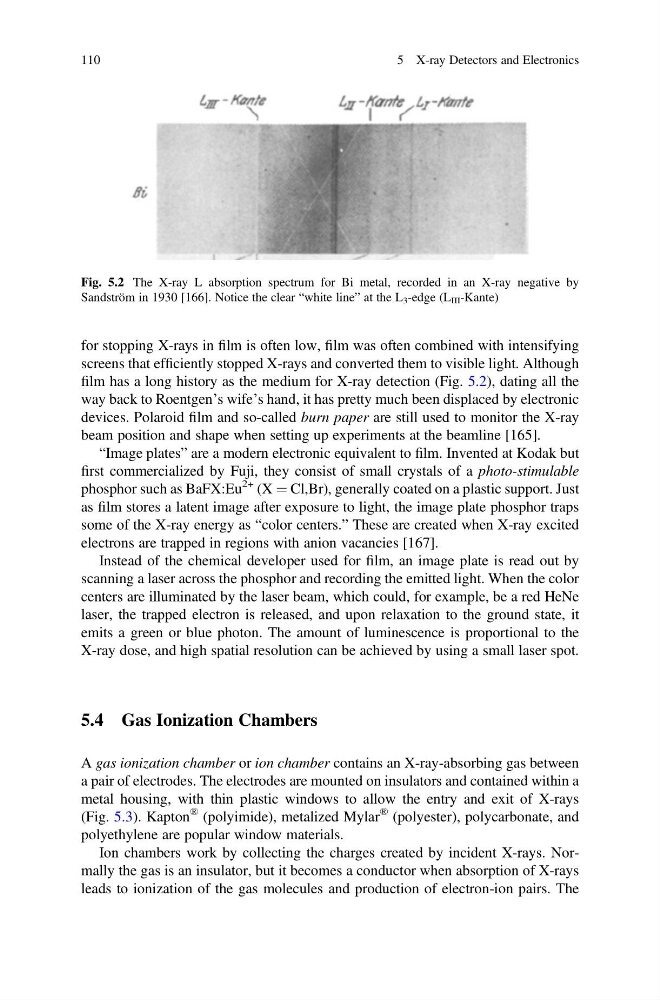
film has a long history as the medium for X-ray detection (Fig. 5.2), dating all the
way back to Roentgen’s wife’s hand, it has pretty much been displaced by electronic
devices. Polaroid film and so-called burn paper are still used to monitor the X-ray
beam position and shape when setting up experiments at the beamline [165].
“Image plates” are a modern electronic equivalent to film. Invented at Kodak but
first commercialized by Fuji, they consist of small crystals of a photo-stimulable
phosphor such as BaFX:Eu
2+ (X ¼ Cl,Br), generally coated on a plastic support. Just
as film stores a latent image after exposure to light, the image plate phosphor traps
some of the X-ray energy as “color centers.” These are created when X-ray excited
electrons are trapped in regions with anion vacancies [167].
Instead of the chemical developer used for film, an image plate is read out by
scanning a laser across the phosphor and recording the emitted light. When the color
centers are illuminated by the laser beam, which could, for example, be a red HeNe
laser, the trapped electron is released, and upon relaxation to the ground state, it
emits a green or blue photon. The amount of luminescence is proportional to the
X-ray dose, and high spatial resolution can be achieved by using a small laser spot.
5.4 Gas Ionization Chambers
A gas ionization chamber or ion chamber contains an X-ray-absorbing gas between
a pair of electrodes. The electrodes are mounted on insulators and contained within a
metal housing, with thin plastic windows to allow the entry and exit of X-rays
(Fig. 5.3). Kapton
® (polyimide), metalized Mylar
® (polyester), polycarbonate, and
polyethylene are popular window materials.
Ion chambers work by collecting the charges created by incident X-rays. Normally the gas is an insulator, but it becomes a conductor when absorption of X-rays
leads to ionization of the gas molecules and production of electron-ion pairs. The
Fig. 5.2 The X-ray L absorption spectrum for Bi metal, recorded in an X-ray negative by
Sandström in 1930 [166]. Notice the clear “white line” at the L 3 -edge (L III -Kante)
110
5 X-ray Detectors and Electronics
screens that efficiently stopped X-rays and converted them to visible light. Although
film has a long history as the medium for X-ray detection (Fig. 5.2), dating all the
way back to Roentgen’s wife’s hand, it has pretty much been displaced by electronic
devices. Polaroid film and so-called burn paper are still used to monitor the X-ray
beam position and shape when setting up experiments at the beamline [165].
“Image plates” are a modern electronic equivalent to film. Invented at Kodak but
first commercialized by Fuji, they consist of small crystals of a photo-stimulable
phosphor such as BaFX:Eu
2+ (X ¼ Cl,Br), generally coated on a plastic support. Just
as film stores a latent image after exposure to light, the image plate phosphor traps
some of the X-ray energy as “color centers.” These are created when X-ray excited
electrons are trapped in regions with anion vacancies [167].
Instead of the chemical developer used for film, an image plate is read out by
scanning a laser across the phosphor and recording the emitted light. When the color
centers are illuminated by the laser beam, which could, for example, be a red HeNe
laser, the trapped electron is released, and upon relaxation to the ground state, it
emits a green or blue photon. The amount of luminescence is proportional to the
X-ray dose, and high spatial resolution can be achieved by using a small laser spot.
5.4 Gas Ionization Chambers
A gas ionization chamber or ion chamber contains an X-ray-absorbing gas between
a pair of electrodes. The electrodes are mounted on insulators and contained within a
metal housing, with thin plastic windows to allow the entry and exit of X-rays
(Fig. 5.3). Kapton
® (polyimide), metalized Mylar
® (polyester), polycarbonate, and
polyethylene are popular window materials.
Ion chambers work by collecting the charges created by incident X-rays. Normally the gas is an insulator, but it becomes a conductor when absorption of X-rays
leads to ionization of the gas molecules and production of electron-ion pairs. The
Fig. 5.2 The X-ray L absorption spectrum for Bi metal, recorded in an X-ray negative by
Sandström in 1930 [166]. Notice the clear “white line” at the L 3 -edge (L III -Kante)
110
5 X-ray Detectors and Electronics