

et al. 2013; McMorran et al. 2014; Kleinschmidt 2015, and references therein). Reflecting their
presumed evolutionary origins, some β-barrel proteins, such as VDAC (Fig. 1.8), are found in the
outer membranes of chloroplasts and mitochondria.
A vast body of data, some of which are presented in Chaps. 6 and 7, suggests that neither
translocons nor a membrane environment is needed for a MP to reach its functional 3D structure,
which is primarily determined by (i) access to an amphipathic medium in which TM segments are
protected from being exposed to water whereas extramembrane ones have access to it and
(ii) interactions of the polypeptide chain with itself. This view is discussed in Popot (2014) and
Popot and Engelman (2016) (for a summary, see Chap. 6, Box 6.3).
1.7.2
Overexpression
Most MPs are not expressed naturally in sufficient amounts for experimental purposes. There are four
approaches to circumventing this problem:
(i) Overexpression in vivo and in situ, the protein being directed toward one of the membrane
compartments of the cell. The main difficulty of this approach is that overexpression of MPs
tends to be toxic and needs to be finely tuned in order not to kill the cells while achieving
decent yields.
(ii) Overexpression in vivo in inclusion bodies (IBs). IBs are nontoxic particles that precipitate
in the cytosol when overexpressed MPs are not targeted to a membrane. IBs permit to
achieve very high yields. However, MPs in IBs are not properly folded. They must be
solubilized under a denatured form in urea or sodium dodecyl sulfate and brought to their
native state, which is rarely straightforward.
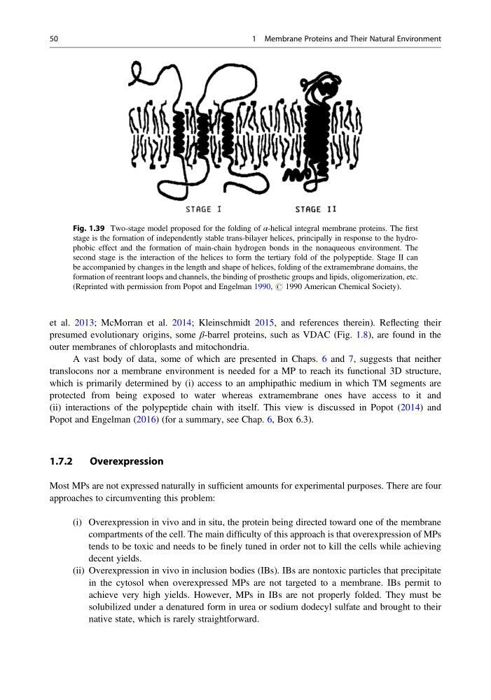
Fig. 1.39 Two-stage model proposed for the folding of α-helical integral membrane proteins. The first
stage is the formation of independently stable trans-bilayer helices, principally in response to the hydrophobic effect and the formation of main-chain hydrogen bonds in the nonaqueous environment. The
second stage is the interaction of the helices to form the tertiary fold of the polypeptide. Stage II can
be accompanied by changes in the length and shape of helices, folding of the extramembrane domains, the
formation of reentrant loops and channels, the binding of prosthetic groups and lipids, oligomerization, etc.
(Reprinted with permission from Popot and Engelman 1990, # 1990 American Chemical Society).
50
1 Membrane Proteins and Their Natural Environment
presumed evolutionary origins, some β-barrel proteins, such as VDAC (Fig. 1.8), are found in the
outer membranes of chloroplasts and mitochondria.
A vast body of data, some of which are presented in Chaps. 6 and 7, suggests that neither
translocons nor a membrane environment is needed for a MP to reach its functional 3D structure,
which is primarily determined by (i) access to an amphipathic medium in which TM segments are
protected from being exposed to water whereas extramembrane ones have access to it and
(ii) interactions of the polypeptide chain with itself. This view is discussed in Popot (2014) and
Popot and Engelman (2016) (for a summary, see Chap. 6, Box 6.3).
1.7.2
Overexpression
Most MPs are not expressed naturally in sufficient amounts for experimental purposes. There are four
approaches to circumventing this problem:
(i) Overexpression in vivo and in situ, the protein being directed toward one of the membrane
compartments of the cell. The main difficulty of this approach is that overexpression of MPs
tends to be toxic and needs to be finely tuned in order not to kill the cells while achieving
decent yields.
(ii) Overexpression in vivo in inclusion bodies (IBs). IBs are nontoxic particles that precipitate
in the cytosol when overexpressed MPs are not targeted to a membrane. IBs permit to
achieve very high yields. However, MPs in IBs are not properly folded. They must be
solubilized under a denatured form in urea or sodium dodecyl sulfate and brought to their
native state, which is rarely straightforward.
Fig. 1.39 Two-stage model proposed for the folding of α-helical integral membrane proteins. The first
stage is the formation of independently stable trans-bilayer helices, principally in response to the hydrophobic effect and the formation of main-chain hydrogen bonds in the nonaqueous environment. The
second stage is the interaction of the helices to form the tertiary fold of the polypeptide. Stage II can
be accompanied by changes in the length and shape of helices, folding of the extramembrane domains, the
formation of reentrant loops and channels, the binding of prosthetic groups and lipids, oligomerization, etc.
(Reprinted with permission from Popot and Engelman 1990, # 1990 American Chemical Society).
50
1 Membrane Proteins and Their Natural Environment