

ESI-LC-MS/MS analysis of tryptic digests following FPOP of both DDM-solubilized and
A8-35-trapped OmpT yielded sequence coverages of 90–95%. Of the 31 predicted tryptic peptides,
13 were found to be modified, with a total of 20 modification sites being identified in both surfactants.
The modified residues were either sulfur-containing or aromatic ones, as expected from their known
propensity to undergo oxidative labeling (Xu and Chance 2007). The oxidation sites detected for
OmpT are the same regardless of the surfactant employed, but the degree of modification of certain
residues varied depending on the protein’s solvent-accessible surface area in each surfactant: some
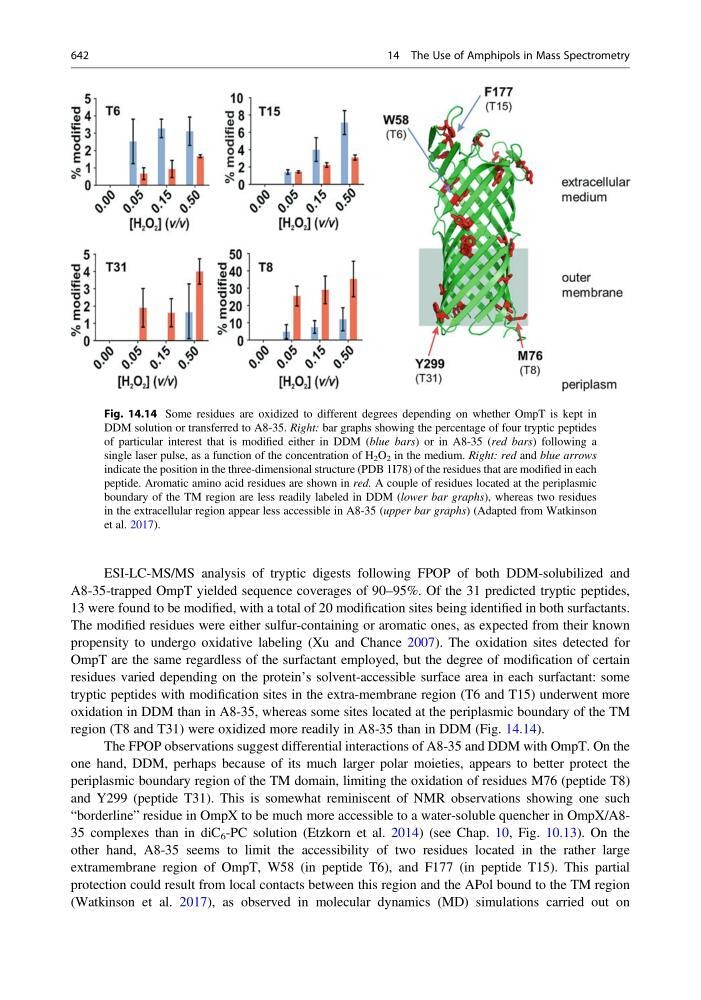
tryptic peptides with modification sites in the extra-membrane region (T6 and T15) underwent more
oxidation in DDM than in A8-35, whereas some sites located at the periplasmic boundary of the TM
region (T8 and T31) were oxidized more readily in A8-35 than in DDM (Fig. 14.14).
The FPOP observations suggest differential interactions of A8-35 and DDM with OmpT. On the
one hand, DDM, perhaps because of its much larger polar moieties, appears to better protect the
periplasmic boundary region of the TM domain, limiting the oxidation of residues M76 (peptide T8)
and Y299 (peptide T31). This is somewhat reminiscent of NMR observations showing one such
“borderline” residue in OmpX to be much more accessible to a water-soluble quencher in OmpX/A835 complexes than in diC 6 -PC solution (Etzkorn et al. 2014) (see Chap. 10, Fig. 10.13). On the
other hand, A8-35 seems to limit the accessibility of two residues located in the rather large
extramembrane region of OmpT, W58 (in peptide T6), and F177 (in peptide T15). This partial
protection could result from local contacts between this region and the APol bound to the TM region
(Watkinson et al. 2017), as observed in molecular dynamics (MD) simulations carried out on
Fig. 14.14 Some residues are oxidized to different degrees depending on whether OmpT is kept in
DDM solution or transferred to A8-35. Right: bar graphs showing the percentage of four tryptic peptides
of particular interest that is modified either in DDM (blue bars) or in A8-35 (red bars) following a
single laser pulse, as a function of the concentration of H 2 O 2 in the medium. Right: red and blue arrows
indicate the position in the three-dimensional structure (PDB 1I78) of the residues that are modified in each
peptide. Aromatic amino acid residues are shown in red. A couple of residues located at the periplasmic
boundary of the TM region are less readily labeled in DDM (lower bar graphs), whereas two residues
in the extracellular region appear less accessible in A8-35 (upper bar graphs) (Adapted from Watkinson
et al. 2017).
642
14 The Use of Amphipols in Mass Spectrometry