

C-termini of each subunit lie in the extracellular space (Fig. 1.26A). Each subunit comprises a large
extracellular region, a TM region comprising four TM helices, labeled M1 to M4, and an intracellular
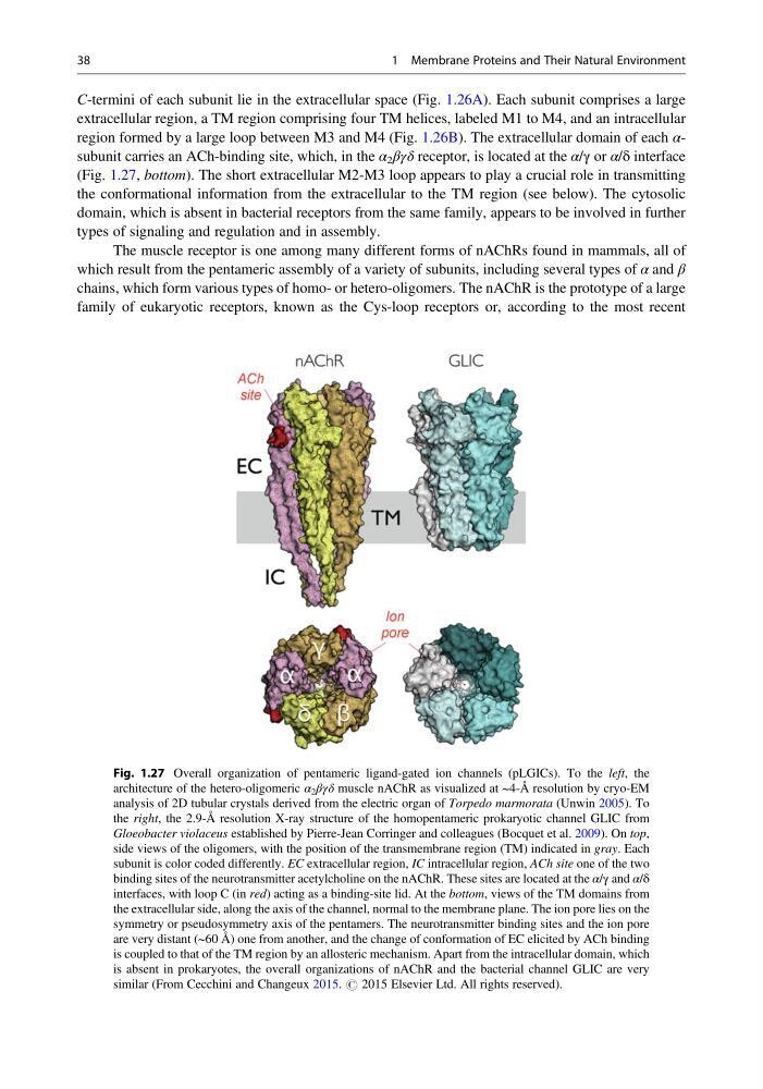
region formed by a large loop between M3 and M4 (Fig. 1.26B). The extracellular domain of each αsubunit carries an ACh-binding site, which, in the α 2 βγδ receptor, is located at the α/γ or α/δ interface
(Fig. 1.27, bottom). The short extracellular M2-M3 loop appears to play a crucial role in transmitting
the conformational information from the extracellular to the TM region (see below). The cytosolic
domain, which is absent in bacterial receptors from the same family, appears to be involved in further
types of signaling and regulation and in assembly.
The muscle receptor is one among many different forms of nAChRs found in mammals, all of
which result from the pentameric assembly of a variety of subunits, including several types of α and β
chains, which form various types of homo- or hetero-oligomers. The nAChR is the prototype of a large
family of eukaryotic receptors, known as the Cys-loop receptors or, according to the most recent
Fig. 1.27 Overall organization of pentameric ligand-gated ion channels (pLGICs). To the left, the
architecture of the hetero-oligomeric α 2 βγδ muscle nAChR as visualized at ~4-Å resolution by cryo-EM
analysis of 2D tubular crystals derived from the electric organ of Torpedo marmorata (Unwin 2005). To
the right, the 2.9-Å resolution X-ray structure of the homopentameric prokaryotic channel GLIC from
Gloeobacter violaceus established by Pierre-Jean Corringer and colleagues (Bocquet et al. 2009). On top,
side views of the oligomers, with the position of the transmembrane region (TM) indicated in gray. Each
subunit is color coded differently. EC extracellular region, IC intracellular region, ACh site one of the two
binding sites of the neurotransmitter acetylcholine on the nAChR. These sites are located at the α/γ and α/δ
interfaces, with loop C (in red) acting as a binding-site lid. At the bottom, views of the TM domains from
the extracellular side, along the axis of the channel, normal to the membrane plane. The ion pore lies on the
symmetry or pseudosymmetry axis of the pentamers. The neurotransmitter binding sites and the ion pore
are very distant (~60 Å) one from another, and the change of conformation of EC elicited by ACh binding
is coupled to that of the TM region by an allosteric mechanism. Apart from the intracellular domain, which
is absent in prokaryotes, the overall organizations of nAChR and the bacterial channel GLIC are very
similar (From Cecchini and Changeux 2015. # 2015 Elsevier Ltd. All rights reserved).
38
1 Membrane Proteins and Their Natural Environment
extracellular region, a TM region comprising four TM helices, labeled M1 to M4, and an intracellular
region formed by a large loop between M3 and M4 (Fig. 1.26B). The extracellular domain of each αsubunit carries an ACh-binding site, which, in the α 2 βγδ receptor, is located at the α/γ or α/δ interface
(Fig. 1.27, bottom). The short extracellular M2-M3 loop appears to play a crucial role in transmitting
the conformational information from the extracellular to the TM region (see below). The cytosolic
domain, which is absent in bacterial receptors from the same family, appears to be involved in further
types of signaling and regulation and in assembly.
The muscle receptor is one among many different forms of nAChRs found in mammals, all of
which result from the pentameric assembly of a variety of subunits, including several types of α and β
chains, which form various types of homo- or hetero-oligomers. The nAChR is the prototype of a large
family of eukaryotic receptors, known as the Cys-loop receptors or, according to the most recent
Fig. 1.27 Overall organization of pentameric ligand-gated ion channels (pLGICs). To the left, the
architecture of the hetero-oligomeric α 2 βγδ muscle nAChR as visualized at ~4-Å resolution by cryo-EM
analysis of 2D tubular crystals derived from the electric organ of Torpedo marmorata (Unwin 2005). To
the right, the 2.9-Å resolution X-ray structure of the homopentameric prokaryotic channel GLIC from
Gloeobacter violaceus established by Pierre-Jean Corringer and colleagues (Bocquet et al. 2009). On top,
side views of the oligomers, with the position of the transmembrane region (TM) indicated in gray. Each
subunit is color coded differently. EC extracellular region, IC intracellular region, ACh site one of the two
binding sites of the neurotransmitter acetylcholine on the nAChR. These sites are located at the α/γ and α/δ
interfaces, with loop C (in red) acting as a binding-site lid. At the bottom, views of the TM domains from
the extracellular side, along the axis of the channel, normal to the membrane plane. The ion pore lies on the
symmetry or pseudosymmetry axis of the pentamers. The neurotransmitter binding sites and the ion pore
are very distant (~60 Å) one from another, and the change of conformation of EC elicited by ACh binding
is coupled to that of the TM region by an allosteric mechanism. Apart from the intracellular domain, which
is absent in prokaryotes, the overall organizations of nAChR and the bacterial channel GLIC are very
similar (From Cecchini and Changeux 2015. # 2015 Elsevier Ltd. All rights reserved).
38
1 Membrane Proteins and Their Natural Environment