

In the absence of ligands, the nAChR is, in majority, in one (or a collection of) resting state(s)
(R), in which the ion channel is closed. R is stabilized by antagonists like curare (hence their
paralyzing effect). According to a model first developed in the laboratory of Jean-Pierre Changeux,
binding of ACh induces a concerted, allosteric conformational change that results in populating the
active state (A), thus opening the TM cation channel and inducing membrane depolarization. Upon
extended application of ACh (hundreds of ms to minutes), two or more further changes of conformation take place, leading to fast or intermediate (I) and to slow (D) desensitized states, in which the
channel is shut. The affinity of the binding sites for ACh increases in the order R < A < I < D, driving
the successive transitions. A number of snake toxins, including α-bungarotoxin (α-Bgt) from the
many-banded krait, Bungarus multicinctus, block access to the ACh-binding sites, paralyzing the
snake’s victims. The binding of α-Bgt is non-covalent but nearly irreversible. The toxin can be labeled
radioactively or fluorescently, making it a remarkable tool for innumerable investigations, examples of
which will be shown in Chaps. 5 and 13. Numerous drugs, including local and general anesthetics,
affect allosterically the receptor’s transitions (see Corringer et al. 2012; Sine 2012; Taly et al. 2014;
Auerbach 2015; Changeux et al. 2015; Nemecz et al. 2016), which are also dependent on the protein’s
membrane environment, in particular on the presence of anionic lipids and cholesterol (reviewed in
Baenziger et al. 2015; Barrantes 2015; Hénault et al. 2015).
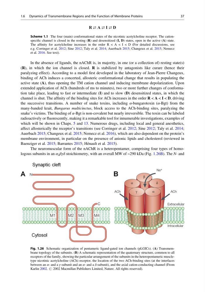
The neuromuscular form of the nAChR is a heteropentamer, comprising four types of homologous subunits in an α 2 βγδ stoichiometry, with an overall MW of ~290 kDa (Fig. 1.26B). The N- and
Fig. 1.26 Schematic organization of pentameric ligand-gated ion channels (pLGICs). (A) Transmembrane topology of the subunits. (B) A schematic representation of the quaternary structure, common to all
receptors of the family, showing the particular arrangement of the subunits in the heteropentameric muscletype nicotinic acetylcholine (ACh) receptor, the location of the two ACh-binding sites (at the interfaces
between an α- and a γ-subunit and an α- and a δ-subunit), and the axial cation-conducting channel (From
Karlin 2002. # 2002 Macmillan Publishers Limited, Nature. All rights reserved).
R A I D
Scheme 1.1 The four (main) conformational states of the nicotinic acetylcholine receptor. The cationspecific channel is closed in the resting (R) and desensitized (I, D) states, open in the active (A) state.
The affinity for acetylcholine increases in the order R < A < I < D (For detailed discussions, see
e.g. Corringer et al. 2012; Sine 2012; Taly et al. 2014; Auerbach 2015; Changeux et al. 2015; Nemecz
et al. 2016. See text).
1.6 Dynamics of Transmembrane Regions and the Function of Membrane Proteins
37
(R), in which the ion channel is closed. R is stabilized by antagonists like curare (hence their
paralyzing effect). According to a model first developed in the laboratory of Jean-Pierre Changeux,
binding of ACh induces a concerted, allosteric conformational change that results in populating the
active state (A), thus opening the TM cation channel and inducing membrane depolarization. Upon
extended application of ACh (hundreds of ms to minutes), two or more further changes of conformation take place, leading to fast or intermediate (I) and to slow (D) desensitized states, in which the
channel is shut. The affinity of the binding sites for ACh increases in the order R < A < I < D, driving
the successive transitions. A number of snake toxins, including α-bungarotoxin (α-Bgt) from the
many-banded krait, Bungarus multicinctus, block access to the ACh-binding sites, paralyzing the
snake’s victims. The binding of α-Bgt is non-covalent but nearly irreversible. The toxin can be labeled
radioactively or fluorescently, making it a remarkable tool for innumerable investigations, examples of
which will be shown in Chaps. 5 and 13. Numerous drugs, including local and general anesthetics,
affect allosterically the receptor’s transitions (see Corringer et al. 2012; Sine 2012; Taly et al. 2014;
Auerbach 2015; Changeux et al. 2015; Nemecz et al. 2016), which are also dependent on the protein’s
membrane environment, in particular on the presence of anionic lipids and cholesterol (reviewed in
Baenziger et al. 2015; Barrantes 2015; Hénault et al. 2015).
The neuromuscular form of the nAChR is a heteropentamer, comprising four types of homologous subunits in an α 2 βγδ stoichiometry, with an overall MW of ~290 kDa (Fig. 1.26B). The N- and
Fig. 1.26 Schematic organization of pentameric ligand-gated ion channels (pLGICs). (A) Transmembrane topology of the subunits. (B) A schematic representation of the quaternary structure, common to all
receptors of the family, showing the particular arrangement of the subunits in the heteropentameric muscletype nicotinic acetylcholine (ACh) receptor, the location of the two ACh-binding sites (at the interfaces
between an α- and a γ-subunit and an α- and a δ-subunit), and the axial cation-conducting channel (From
Karlin 2002. # 2002 Macmillan Publishers Limited, Nature. All rights reserved).
R A I D
Scheme 1.1 The four (main) conformational states of the nicotinic acetylcholine receptor. The cationspecific channel is closed in the resting (R) and desensitized (I, D) states, open in the active (A) state.
The affinity for acetylcholine increases in the order R < A < I < D (For detailed discussions, see
e.g. Corringer et al. 2012; Sine 2012; Taly et al. 2014; Auerbach 2015; Changeux et al. 2015; Nemecz
et al. 2016. See text).
1.6 Dynamics of Transmembrane Regions and the Function of Membrane Proteins
37