

It is notable that, for this particular study, PMAL-C8 provided better data than A8-35 (Y. Cheng,
personal communication), perhaps because of its lower net charge density, illustrating once more that
the concept of “one-fits-all” does not work with MPs.
A brief mention should be made of Study 12.20 (Chowdhury et al. 2015). It does not bear on a
MP or MP complex, but on dynactin, a cytosolic protein involved in the transport of cellular cargoes by
microtubules. The cryo-EM protocol used mentions that purified dynactin was applied to freshly glowdischarged holey grids. Immediately before application of the protein sample on the grid, a dilute
(0.25 gÁL
À1 ) solution of A8-35 was mixed with the sample to aid in dispersing dynactin particles across
the holes in the carbon film. Excess sample was manually blotted with filter paper for ~5 to 7 s, and
the sample was immediately vitrified by plunge-freezing in liquid-ethane slurry at À179
C. These
observations are interesting, because it seems unlikely that A8-35 bind to dynactin. Therefore, the
better dispersion of the particles, which reminds one of the observations made with Complex I
(Flötenmeyer et al. 2007; see Fig. 12.9), the ryanodine receptor (Baker et al. 2015; see below,
Fig. 12.27), and an engineered AcrAB-TolC complex (Jeong et al. 2016), is probably related to either
the thickness of the film or its surface properties (see below, § 12.3.4.2). Similarly, A8-35 was used in
the cryo-EM study of a (non-membrane) nuclease as a substitute to detergent, in order to make the
holey carbon film hydrophilic (Wilkinson et al. 2016a, b). It is indeed not unusual for microscopists
working on non-membrane objects to add some detergent at submicellar concentrations to their
samples in order to modify the distribution or orientation of the particles (for a recent discussion of
the formation and effects of air/water surfactant monolayers in cryo-EM, see Glaeser et al. 2016).
Finally, it is worth mentioning observations reported in Study 12.19, a review of cryo-EM
studies of the ryanodine receptor (RyR) (Baker et al. 2015) written shortly before the structure of this
huge, homotetrameric complex (~2.3 MDa) was solved to 3.8 Å (Yan et al. 2015) and 4.8 Å (Zalk et al.
2015) by cryo-EM studies in detergent solutions. The review presents original data collected on Type 1
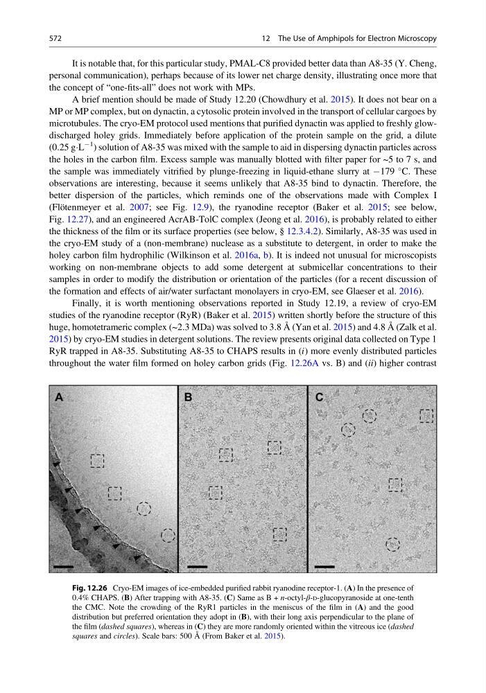
RyR trapped in A8-35. Substituting A8-35 to CHAPS results in (i) more evenly distributed particles
throughout the water film formed on holey carbon grids (Fig. 12.26A vs. B) and (ii) higher contrast
Fig. 12.26 Cryo-EM images of ice-embedded purified rabbit ryanodine receptor-1. (A) In the presence of
0.4% CHAPS. (B) After trapping with A8-35. (C) Same as B + n-octyl-β-D-glucopyranoside at one-tenth
the CMC. Note the crowding of the RyR1 particles in the meniscus of the film in (A) and the good
distribution but preferred orientation they adopt in (B), with their long axis perpendicular to the plane of
the film (dashed squares), whereas in (C) they are more randomly oriented within the vitreous ice (dashed
squares and circles). Scale bars: 500 Å (From Baker et al. 2015).
572
12 The Use of Amphipols for Electron Microscopy
personal communication), perhaps because of its lower net charge density, illustrating once more that
the concept of “one-fits-all” does not work with MPs.
A brief mention should be made of Study 12.20 (Chowdhury et al. 2015). It does not bear on a
MP or MP complex, but on dynactin, a cytosolic protein involved in the transport of cellular cargoes by
microtubules. The cryo-EM protocol used mentions that purified dynactin was applied to freshly glowdischarged holey grids. Immediately before application of the protein sample on the grid, a dilute
(0.25 gÁL
À1 ) solution of A8-35 was mixed with the sample to aid in dispersing dynactin particles across
the holes in the carbon film. Excess sample was manually blotted with filter paper for ~5 to 7 s, and
the sample was immediately vitrified by plunge-freezing in liquid-ethane slurry at À179
C. These
observations are interesting, because it seems unlikely that A8-35 bind to dynactin. Therefore, the
better dispersion of the particles, which reminds one of the observations made with Complex I
(Flötenmeyer et al. 2007; see Fig. 12.9), the ryanodine receptor (Baker et al. 2015; see below,
Fig. 12.27), and an engineered AcrAB-TolC complex (Jeong et al. 2016), is probably related to either
the thickness of the film or its surface properties (see below, § 12.3.4.2). Similarly, A8-35 was used in
the cryo-EM study of a (non-membrane) nuclease as a substitute to detergent, in order to make the
holey carbon film hydrophilic (Wilkinson et al. 2016a, b). It is indeed not unusual for microscopists
working on non-membrane objects to add some detergent at submicellar concentrations to their
samples in order to modify the distribution or orientation of the particles (for a recent discussion of
the formation and effects of air/water surfactant monolayers in cryo-EM, see Glaeser et al. 2016).
Finally, it is worth mentioning observations reported in Study 12.19, a review of cryo-EM
studies of the ryanodine receptor (RyR) (Baker et al. 2015) written shortly before the structure of this
huge, homotetrameric complex (~2.3 MDa) was solved to 3.8 Å (Yan et al. 2015) and 4.8 Å (Zalk et al.
2015) by cryo-EM studies in detergent solutions. The review presents original data collected on Type 1
RyR trapped in A8-35. Substituting A8-35 to CHAPS results in (i) more evenly distributed particles
throughout the water film formed on holey carbon grids (Fig. 12.26A vs. B) and (ii) higher contrast
Fig. 12.26 Cryo-EM images of ice-embedded purified rabbit ryanodine receptor-1. (A) In the presence of
0.4% CHAPS. (B) After trapping with A8-35. (C) Same as B + n-octyl-β-D-glucopyranoside at one-tenth
the CMC. Note the crowding of the RyR1 particles in the meniscus of the film in (A) and the good
distribution but preferred orientation they adopt in (B), with their long axis perpendicular to the plane of
the film (dashed squares), whereas in (C) they are more randomly oriented within the vitreous ice (dashed
squares and circles). Scale bars: 500 Å (From Baker et al. 2015).
572
12 The Use of Amphipols for Electron Microscopy