

Before moving on, it is useful to say a word of which types of MP crystals could be a priori
expected. Those were distinguished early on by Hartmut Michel in an insightful mini-review (Michel
1983; for a more recent, extensive analysis of the organization of the protein and detergent in MP
crystals, see Schulz 2011). H. Michel described two types of 3D crystals according to the kinds of
contacts the proteins would make between themselves and with their environment. Type I crystals
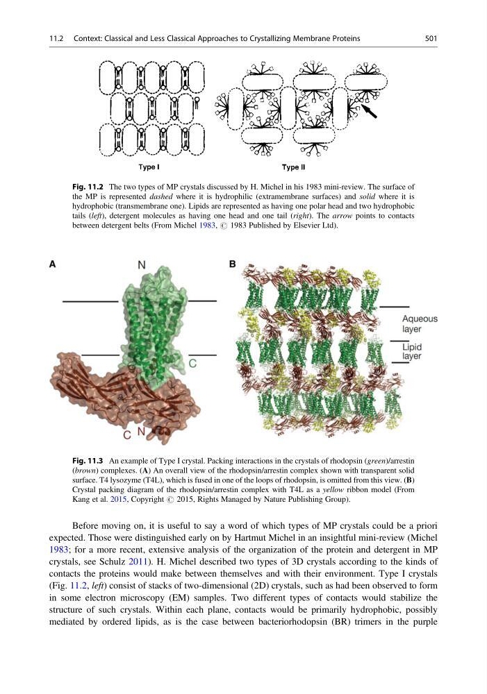
(Fig. 11.2, left) consist of stacks of two-dimensional (2D) crystals, such as had been observed to form
in some electron microscopy (EM) samples. Two different types of contacts would stabilize the
structure of such crystals. Within each plane, contacts would be primarily hydrophobic, possibly
mediated by ordered lipids, as is the case between bacteriorhodopsin (BR) trimers in the purple
Fig. 11.2 The two types of MP crystals discussed by H. Michel in his 1983 mini-review. The surface of
the MP is represented dashed where it is hydrophilic (extramembrane surfaces) and solid where it is
hydrophobic (transmembrane one). Lipids are represented as having one polar head and two hydrophobic
tails (left), detergent molecules as having one head and one tail (right). The arrow points to contacts
between detergent belts (From Michel 1983, # 1983 Published by Elsevier Ltd).
Fig. 11.3 An example of Type I crystal. Packing interactions in the crystals of rhodopsin (green)/arrestin
(brown) complexes. (A) An overall view of the rhodopsin/arrestin complex shown with transparent solid
surface. T4 lysozyme (T4L), which is fused in one of the loops of rhodopsin, is omitted from this view. (B)
Crystal packing diagram of the rhodopsin/arrestin complex with T4L as a yellow ribbon model (From
Kang et al. 2015, Copyright # 2015, Rights Managed by Nature Publishing Group).
11.2 Context: Classical and Less Classical Approaches to Crystallizing Membrane Proteins
501
expected. Those were distinguished early on by Hartmut Michel in an insightful mini-review (Michel
1983; for a more recent, extensive analysis of the organization of the protein and detergent in MP
crystals, see Schulz 2011). H. Michel described two types of 3D crystals according to the kinds of
contacts the proteins would make between themselves and with their environment. Type I crystals
(Fig. 11.2, left) consist of stacks of two-dimensional (2D) crystals, such as had been observed to form
in some electron microscopy (EM) samples. Two different types of contacts would stabilize the
structure of such crystals. Within each plane, contacts would be primarily hydrophobic, possibly
mediated by ordered lipids, as is the case between bacteriorhodopsin (BR) trimers in the purple
Fig. 11.2 The two types of MP crystals discussed by H. Michel in his 1983 mini-review. The surface of
the MP is represented dashed where it is hydrophilic (extramembrane surfaces) and solid where it is
hydrophobic (transmembrane one). Lipids are represented as having one polar head and two hydrophobic
tails (left), detergent molecules as having one head and one tail (right). The arrow points to contacts
between detergent belts (From Michel 1983, # 1983 Published by Elsevier Ltd).
Fig. 11.3 An example of Type I crystal. Packing interactions in the crystals of rhodopsin (green)/arrestin
(brown) complexes. (A) An overall view of the rhodopsin/arrestin complex shown with transparent solid
surface. T4 lysozyme (T4L), which is fused in one of the loops of rhodopsin, is omitted from this view. (B)
Crystal packing diagram of the rhodopsin/arrestin complex with T4L as a yellow ribbon model (From
Kang et al. 2015, Copyright # 2015, Rights Managed by Nature Publishing Group).
11.2 Context: Classical and Less Classical Approaches to Crystallizing Membrane Proteins
501