

structure adopted by ligands while bound to them is a major goal of pharmacological research. APols
have proven to be an efficient medium into which fold GPCRs from a denatured state, some of which
had never been folded to any appreciable extent using more classical approaches (Dahmane et al. 2009;
Banères et al. 2011; Bazzacco et al. 2012; see Chap. 6). The low-affinity leukotriene receptor (BLT2)
is involved in inflammatory diseases and allergic responses. BLT2 is activated by leukotriene B 4
(5S,12R-dihydroxy-6Z,8E,10E,14Z-eicosatetraenoic acid; LTB 4 ), a member of the family of
eicosanoids. Before its APol-assisted folding was developed (Dahmane et al. 2009; see Chap. 6,
§ 6.3.1.1.2), BLT2 had never been obtained in amounts suitable for structural studies, and the structure
of LTB 4 bound to BLT2 was unknown. LTB 4 is characterized by a triene motif and the presence of two
hydroxyl groups and an acidic function (Fig. 10.23). The presence of conjugated and unconjugated
double bonds and that of hydroxyl and carboxyl groups creates a wide variety of chemical environment, resulting in a broad range of
1 H chemical shifts, which is a great asset in establishing an NMR
structure.
In Study 10.4 (Table 10.1; Catoire et al. 2010a), BLT2 was expressed in perdeuterated,
15 Nlabeled form in E. coli inclusion bodies, solubilized in SDS, folded in DAPol, and the structure of
BLT2-bound LTB 4 deduced from intramolecular transferred NOE signals. In order to separate the
130
9.7 9.5
Y71
Y87
Y109
Q61
M21
Q11
Q17
Q114
N19
N74
N34
N25+
Y127
S54
S35
M118
N116
S126
S42
G38
A67
E119
E32
Y30
F115
F24
F125
Y28
Y62
Y98
Y80+
A111
Y146
K20
K27
Q129
F43
F148
A14
A122
Y45
V135
V121
V39
K99
?
?
?
?
?
?
?
V5
V137
V85
V83
V82
G86
G41
G22
G138
G84
G106
G8
G68
G110 G7 G64
G145
G81
G112
G143
G23
N120
V144
T44
T4
T6
T66
T46
T93
W76
D75
S78
E47 T97
T139
D13
D33
S12
S49
A142
R133
R50
L26
L123
L113
R29
R131
R147
A10
A77
A70
D56
S108
L37
R72
I132
I73
E31 D136
D124 W140
I40
I65
I141
I79
9.3 9.1 8.9 8.7 8.5
1 H (ppm)
15
N (ppm)
8.3 8.1 7.9 7.7 7.5 7.3 7.1
128
126
124
122
120
118
116
114
112
110
108
106
104
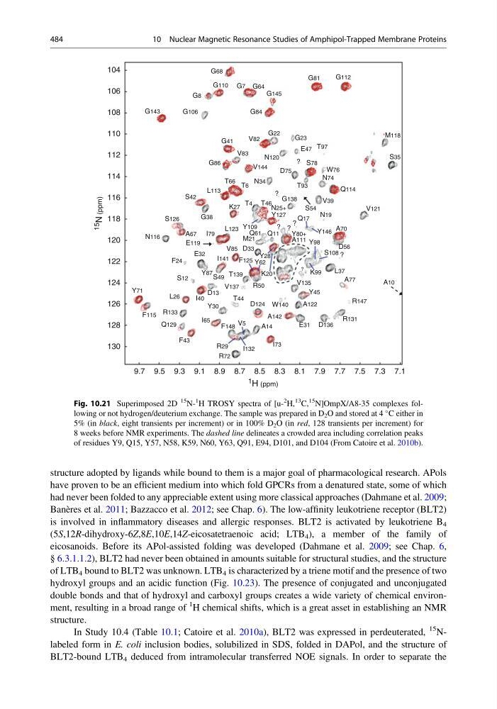
Fig. 10.21 Superimposed 2D
15
N1
H TROSY spectra of [u2
H,
13
C,
15
N]OmpX/A8-35 complexes following or not hydrogen/deuterium exchange. The sample was prepared in D 2 O and stored at 4
C either in
5% (in black, eight transients per increment) or in 100% D 2 O (in red, 128 transients per increment) for
8 weeks before NMR experiments. The dashed line delineates a crowded area including correlation peaks
of residues Y9, Q15, Y57, N58, K59, N60, Y63, Q91, E94, D101, and D104 (From Catoire et al. 2010b).
484
10 Nuclear Magnetic Resonance Studies of Amphipol-Trapped Membrane Proteins
have proven to be an efficient medium into which fold GPCRs from a denatured state, some of which
had never been folded to any appreciable extent using more classical approaches (Dahmane et al. 2009;
Banères et al. 2011; Bazzacco et al. 2012; see Chap. 6). The low-affinity leukotriene receptor (BLT2)
is involved in inflammatory diseases and allergic responses. BLT2 is activated by leukotriene B 4
(5S,12R-dihydroxy-6Z,8E,10E,14Z-eicosatetraenoic acid; LTB 4 ), a member of the family of
eicosanoids. Before its APol-assisted folding was developed (Dahmane et al. 2009; see Chap. 6,
§ 6.3.1.1.2), BLT2 had never been obtained in amounts suitable for structural studies, and the structure
of LTB 4 bound to BLT2 was unknown. LTB 4 is characterized by a triene motif and the presence of two
hydroxyl groups and an acidic function (Fig. 10.23). The presence of conjugated and unconjugated
double bonds and that of hydroxyl and carboxyl groups creates a wide variety of chemical environment, resulting in a broad range of
1 H chemical shifts, which is a great asset in establishing an NMR
structure.
In Study 10.4 (Table 10.1; Catoire et al. 2010a), BLT2 was expressed in perdeuterated,
15 Nlabeled form in E. coli inclusion bodies, solubilized in SDS, folded in DAPol, and the structure of
BLT2-bound LTB 4 deduced from intramolecular transferred NOE signals. In order to separate the
130
9.7 9.5
Y71
Y87
Y109
Q61
M21
Q11
Q17
Q114
N19
N74
N34
N25+
Y127
S54
S35
M118
N116
S126
S42
G38
A67
E119
E32
Y30
F115
F24
F125
Y28
Y62
Y98
Y80+
A111
Y146
K20
K27
Q129
F43
F148
A14
A122
Y45
V135
V121
V39
K99
?
?
?
?
?
?
?
V5
V137
V85
V83
V82
G86
G41
G22
G138
G84
G106
G8
G68
G110 G7 G64
G145
G81
G112
G143
G23
N120
V144
T44
T4
T6
T66
T46
T93
W76
D75
S78
E47 T97
T139
D13
D33
S12
S49
A142
R133
R50
L26
L123
L113
R29
R131
R147
A10
A77
A70
D56
S108
L37
R72
I132
I73
E31 D136
D124 W140
I40
I65
I141
I79
9.3 9.1 8.9 8.7 8.5
1 H (ppm)
15
N (ppm)
8.3 8.1 7.9 7.7 7.5 7.3 7.1
128
126
124
122
120
118
116
114
112
110
108
106
104
Fig. 10.21 Superimposed 2D
15
N1
H TROSY spectra of [u2
H,
13
C,
15
N]OmpX/A8-35 complexes following or not hydrogen/deuterium exchange. The sample was prepared in D 2 O and stored at 4
C either in
5% (in black, eight transients per increment) or in 100% D 2 O (in red, 128 transients per increment) for
8 weeks before NMR experiments. The dashed line delineates a crowded area including correlation peaks
of residues Y9, Q15, Y57, N58, K59, N60, Y63, Q91, E94, D101, and D104 (From Catoire et al. 2010b).
484
10 Nuclear Magnetic Resonance Studies of Amphipol-Trapped Membrane Proteins