

10.3.1.2 Mapping Membrane Protein/Amphipol Interactions by NMR
As described in Chap. 5, a consistent picture of the composition, organization, and dynamics of
MP/APol complexes has emerged from an ensemble of biochemical and biophysical studies including
small-angle radiation scattering, NMR, EM, and mass spectrometry data, as well as MD calculations.
NMR has been particularly instrumental in mapping MP/APol interactions and the accessibility to the
solvent of the surface of APol-trapped proteins.
As noted above, Study 10.1 (Table 10.1) exploited differences between
1 H15 N TROSY-HSQC
spectra of tOmpA collected either in unlabeled A8-35 (HAPol) or in DAPol to map the distribution of
the polymer at the surface of the protein. The broadening of the
1 H resonance lines of a subset of
1 H
N
nuclei was indeed observed to correlate with their position in the 3D structure. This information is
patchy, because less than half of the amide protons of tOmpA are assigned (colored residues in
Fig. 10.11), but it nevertheless provided a first atomic-level description of the way an APol interacts
with the MP it keeps soluble. In brief, two sets of
1 H15 N TROSY-HSQC spectra were recorded at high
magnetic field (ν H ¼ 800 MHz), one with HAPol- and the other with DAPol-trapped [u2 H,
15 N]tOmpA.
Half-height linewidths were compared in the
1 H dimension, because the coupling with remote protons is
more pronounced with
1 H than with
15 N nuclei (Pervushin et al. 1997). The extent of relative
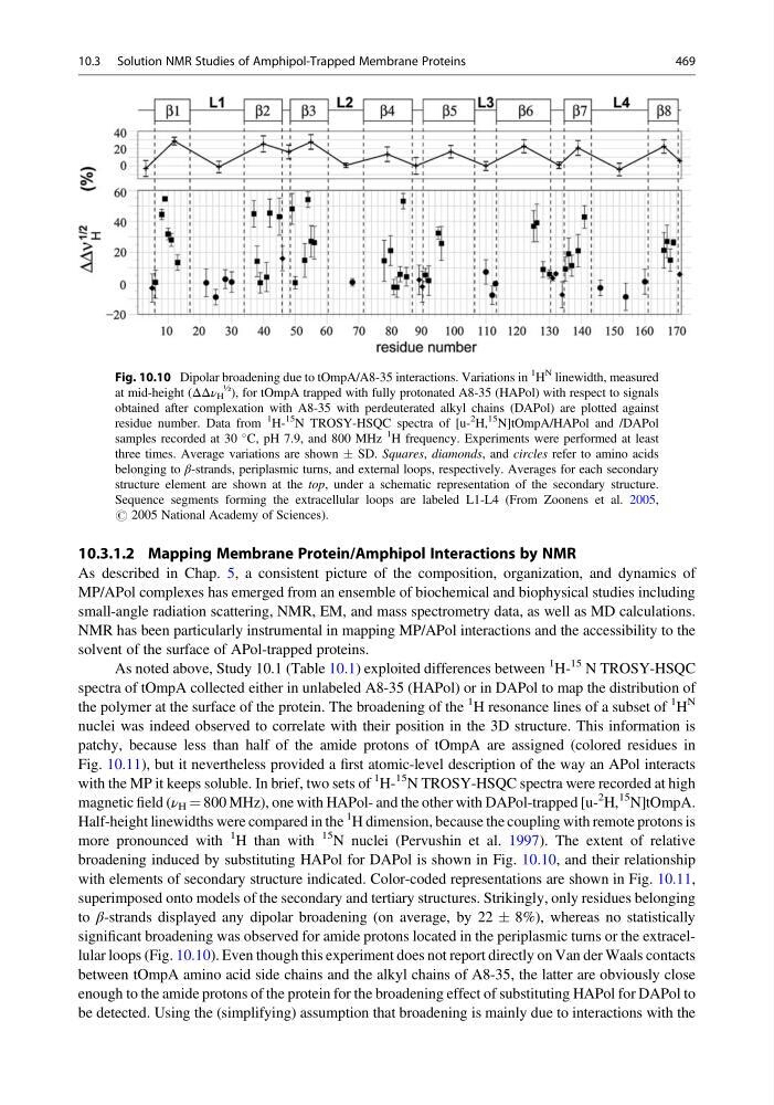
broadening induced by substituting HAPol for DAPol is shown in Fig. 10.10, and their relationship
with elements of secondary structure indicated. Color-coded representations are shown in Fig. 10.11,
superimposed onto models of the secondary and tertiary structures. Strikingly, only residues belonging
to β-strands displayed any dipolar broadening (on average, by 22 Æ 8%), whereas no statistically
significant broadening was observed for amide protons located in the periplasmic turns or the extracellular loops (Fig. 10.10). Even though this experiment does not report directly on Van der Waals contacts
between tOmpA amino acid side chains and the alkyl chains of A8-35, the latter are obviously close
enough to the amide protons of the protein for the broadening effect of substituting HAPol for DAPol to
be detected. Using the (simplifying) assumption that broadening is mainly due to interactions with the
Fig. 10.10 Dipolar broadening due to tOmpA/A8-35 interactions. Variations in
1
H
N linewidth, measured
at mid-height (ΔΔν H
½
), for tOmpA trapped with fully protonated A8-35 (HAPol) with respect to signals
obtained after complexation with A8-35 with perdeuterated alkyl chains (DAPol) are plotted against
residue number. Data from
1
H15
N TROSY-HSQC spectra of [u2
H,
15 N]tOmpA/HAPol and /DAPol
samples recorded at 30
C, pH 7.9, and 800 MHz
1
H frequency. Experiments were performed at least
three times. Average variations are shown Æ SD. Squares, diamonds, and circles refer to amino acids
belonging to β-strands, periplasmic turns, and external loops, respectively. Averages for each secondary
structure element are shown at the top, under a schematic representation of the secondary structure.
Sequence segments forming the extracellular loops are labeled L1-L4 (From Zoonens et al. 2005,
# 2005 National Academy of Sciences).
10.3 Solution NMR Studies of Amphipol-Trapped Membrane Proteins
469
As described in Chap. 5, a consistent picture of the composition, organization, and dynamics of
MP/APol complexes has emerged from an ensemble of biochemical and biophysical studies including
small-angle radiation scattering, NMR, EM, and mass spectrometry data, as well as MD calculations.
NMR has been particularly instrumental in mapping MP/APol interactions and the accessibility to the
solvent of the surface of APol-trapped proteins.
As noted above, Study 10.1 (Table 10.1) exploited differences between
1 H15 N TROSY-HSQC
spectra of tOmpA collected either in unlabeled A8-35 (HAPol) or in DAPol to map the distribution of
the polymer at the surface of the protein. The broadening of the
1 H resonance lines of a subset of
1 H
N
nuclei was indeed observed to correlate with their position in the 3D structure. This information is
patchy, because less than half of the amide protons of tOmpA are assigned (colored residues in
Fig. 10.11), but it nevertheless provided a first atomic-level description of the way an APol interacts
with the MP it keeps soluble. In brief, two sets of
1 H15 N TROSY-HSQC spectra were recorded at high
magnetic field (ν H ¼ 800 MHz), one with HAPol- and the other with DAPol-trapped [u2 H,
15 N]tOmpA.
Half-height linewidths were compared in the
1 H dimension, because the coupling with remote protons is
more pronounced with
1 H than with
15 N nuclei (Pervushin et al. 1997). The extent of relative
broadening induced by substituting HAPol for DAPol is shown in Fig. 10.10, and their relationship
with elements of secondary structure indicated. Color-coded representations are shown in Fig. 10.11,
superimposed onto models of the secondary and tertiary structures. Strikingly, only residues belonging
to β-strands displayed any dipolar broadening (on average, by 22 Æ 8%), whereas no statistically
significant broadening was observed for amide protons located in the periplasmic turns or the extracellular loops (Fig. 10.10). Even though this experiment does not report directly on Van der Waals contacts
between tOmpA amino acid side chains and the alkyl chains of A8-35, the latter are obviously close
enough to the amide protons of the protein for the broadening effect of substituting HAPol for DAPol to
be detected. Using the (simplifying) assumption that broadening is mainly due to interactions with the
Fig. 10.10 Dipolar broadening due to tOmpA/A8-35 interactions. Variations in
1
H
N linewidth, measured
at mid-height (ΔΔν H
½
), for tOmpA trapped with fully protonated A8-35 (HAPol) with respect to signals
obtained after complexation with A8-35 with perdeuterated alkyl chains (DAPol) are plotted against
residue number. Data from
1
H15
N TROSY-HSQC spectra of [u2
H,
15 N]tOmpA/HAPol and /DAPol
samples recorded at 30
C, pH 7.9, and 800 MHz
1
H frequency. Experiments were performed at least
three times. Average variations are shown Æ SD. Squares, diamonds, and circles refer to amino acids
belonging to β-strands, periplasmic turns, and external loops, respectively. Averages for each secondary
structure element are shown at the top, under a schematic representation of the secondary structure.
Sequence segments forming the extracellular loops are labeled L1-L4 (From Zoonens et al. 2005,
# 2005 National Academy of Sciences).
10.3 Solution NMR Studies of Amphipol-Trapped Membrane Proteins
469