

acquired at 560 nm, so as to focus on the distribution of native BR. In D 2
18 O buffer, the M b for
BR/HAPol complexes is close to null, the particles being only slightly redistributed even at the highest
angular velocity (Fig. 9.13C). Despite the slight heterogeneity of this sample (~9% dimer according to
sedimentation velocity data), rather good fits were obtained assuming a single species of
non-interacting particles (Fig. 9.13A–C). Two other samples of BR/HAPol and BR/DAPol were
investigated in H 2 O and D 2 O buffers, using longer columns. Plots of M b values from all equilibrium
sedimentation experiments as a function of solvent density indicate a good consistency of the results
(Fig. 9.13D, E).
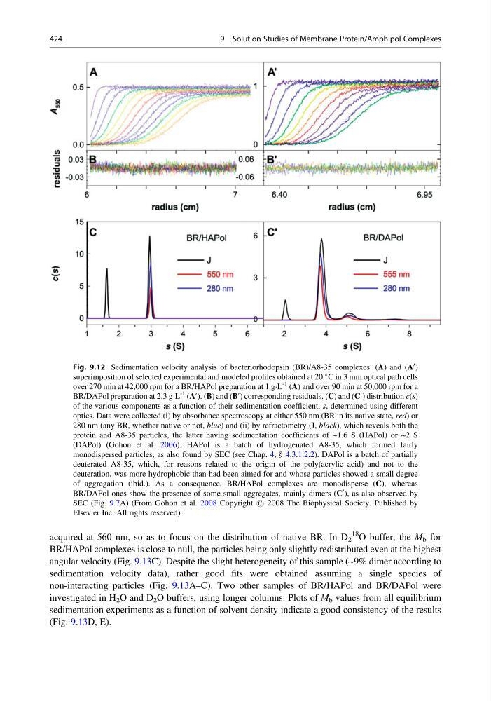
Fig. 9.12 Sedimentation velocity analysis of bacteriorhodopsin (BR)/A8-35 complexes. (A) and (A
0 )
superimposition of selected experimental and modeled profiles obtained at 20
C in 3 mm optical path cells
over 270 min at 42,000 rpm for a BR/HAPol preparation at 1 gÁL
-1 (A) and over 90 min at 50,000 rpm for a
BR/DAPol preparation at 2.3 gÁL
-1 (A
0 ). (B) and (B
0 ) corresponding residuals. (C) and (C
0 ) distribution c(s)
of the various components as a function of their sedimentation coefficient, s, determined using different
optics. Data were collected (i) by absorbance spectroscopy at either 550 nm (BR in its native state, red) or
280 nm (any BR, whether native or not, blue) and (ii) by refractometry (J, black), which reveals both the
protein and A8-35 particles, the latter having sedimentation coefficients of ~1.6 S (HAPol) or ~2 S
(DAPol) (Gohon et al. 2006). HAPol is a batch of hydrogenated A8-35, which formed fairly
monodispersed particles, as also found by SEC (see Chap. 4, § 4.3.1.2.2). DAPol is a batch of partially
deuterated A8-35, which, for reasons related to the origin of the poly(acrylic acid) and not to the
deuteration, was more hydrophobic than had been aimed for and whose particles showed a small degree
of aggregation (ibid.). As a consequence, BR/HAPol complexes are monodisperse (C), whereas
BR/DAPol ones show the presence of some small aggregates, mainly dimers (C
0 ), as also observed by
SEC (Fig. 9.7A) (From Gohon et al. 2008 Copyright # 2008 The Biophysical Society. Published by
Elsevier Inc. All rights reserved).
424
9 Solution Studies of Membrane Protein/Amphipol Complexes
18 O buffer, the M b for
BR/HAPol complexes is close to null, the particles being only slightly redistributed even at the highest
angular velocity (Fig. 9.13C). Despite the slight heterogeneity of this sample (~9% dimer according to
sedimentation velocity data), rather good fits were obtained assuming a single species of
non-interacting particles (Fig. 9.13A–C). Two other samples of BR/HAPol and BR/DAPol were
investigated in H 2 O and D 2 O buffers, using longer columns. Plots of M b values from all equilibrium
sedimentation experiments as a function of solvent density indicate a good consistency of the results
(Fig. 9.13D, E).
Fig. 9.12 Sedimentation velocity analysis of bacteriorhodopsin (BR)/A8-35 complexes. (A) and (A
0 )
superimposition of selected experimental and modeled profiles obtained at 20
C in 3 mm optical path cells
over 270 min at 42,000 rpm for a BR/HAPol preparation at 1 gÁL
-1 (A) and over 90 min at 50,000 rpm for a
BR/DAPol preparation at 2.3 gÁL
-1 (A
0 ). (B) and (B
0 ) corresponding residuals. (C) and (C
0 ) distribution c(s)
of the various components as a function of their sedimentation coefficient, s, determined using different
optics. Data were collected (i) by absorbance spectroscopy at either 550 nm (BR in its native state, red) or
280 nm (any BR, whether native or not, blue) and (ii) by refractometry (J, black), which reveals both the
protein and A8-35 particles, the latter having sedimentation coefficients of ~1.6 S (HAPol) or ~2 S
(DAPol) (Gohon et al. 2006). HAPol is a batch of hydrogenated A8-35, which formed fairly
monodispersed particles, as also found by SEC (see Chap. 4, § 4.3.1.2.2). DAPol is a batch of partially
deuterated A8-35, which, for reasons related to the origin of the poly(acrylic acid) and not to the
deuteration, was more hydrophobic than had been aimed for and whose particles showed a small degree
of aggregation (ibid.). As a consequence, BR/HAPol complexes are monodisperse (C), whereas
BR/DAPol ones show the presence of some small aggregates, mainly dimers (C
0 ), as also observed by
SEC (Fig. 9.7A) (From Gohon et al. 2008 Copyright # 2008 The Biophysical Society. Published by
Elsevier Inc. All rights reserved).
424
9 Solution Studies of Membrane Protein/Amphipol Complexes