

9.1
Introduction
At the onset of any structural or functional study of an amphipol (APol)-trapped membrane protein
(MP), it is advisable to check on the size and homogeneity of the complexes. It is, indeed, very useful
and often essential to know whether one is working with monodisperse, homogeneous monomers or
oligomers, with a mixture thereof, or with partially aggregated preparations. The interpretation of
radiation scattering data, for instance, critically depends on the association state of the protein. Many
types of solution studies can give access to the mass, shape, composition, homogeneity, and interparticle interactions of the complexes, and some of them can be used to gather information about their
internal organization, such as the arrangement of subunits with respect to one another or to the
transmembrane (TM) region, the disposition of the protein, lipid and APol components relative to
each other, etc.
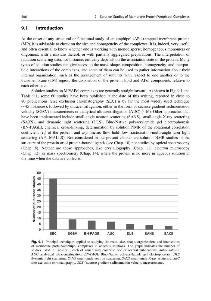
Solution studies on MP/APol complexes are generally straightforward. As shown in Fig. 9.1 and
Table 9.1, some 60 studies have been published at the date of this writing, reported in close to
80 publications. Size exclusion chromatography (SEC) is by far the most widely used technique
(~45 instances), followed by ultracentrifugation, either in the form of sucrose gradient sedimentation
velocity (SGSV) measurements or analytical ultracentrifugation (AUC) (~16). Other approaches that
have been implemented include small-angle neutron scattering (SANS), small-angle X-ray scattering
(SAXS), and dynamic light scattering (DLS), Blue-Native polyacrylamide gel electrophoresis
(BN-PAGE), chemical cross-linking, determination by solution NMR of the rotational correlation
coefficient (τ c ) of the protein, and asymmetric flow field-flow fractionation-multi-angle laser light
scattering (AF4-MALLS). Not considered in the present chapter are solution NMR studies of the
structure of the protein or of protein-bound ligands (see Chap. 10) nor studies by optical spectroscopy
(Chap. 8). Neither are those approaches, like crystallography (Chap. 11), electron microscopy
(Chap. 12), or mass spectrometry (Chap. 14), where the protein is no more in aqueous solution at
the time when the data are collected.
Fig. 9.1 Principal techniques applied to studying the mass, size, shape, organization, and interactions
of membrane protein/amphipol complexes in aqueous solutions. The graph indicates the number of
studies listed in Table 9.1, each of which may comprise one or several publications. Abbreviations:
AUC analytical ultracentrifugation, BN-PAGE Blue-Native polyacrylamide gel electrophoresis, DLS
dynamic light scattering, SANS small-angle neutron scattering, SAXS small-angle X-ray scattering, SEC
size exclusion chromatography, SGSV sucrose gradient sedimentation velocity measurements.
406
9 Solution Studies of Membrane Protein/Amphipol Complexes
Introduction
At the onset of any structural or functional study of an amphipol (APol)-trapped membrane protein
(MP), it is advisable to check on the size and homogeneity of the complexes. It is, indeed, very useful
and often essential to know whether one is working with monodisperse, homogeneous monomers or
oligomers, with a mixture thereof, or with partially aggregated preparations. The interpretation of
radiation scattering data, for instance, critically depends on the association state of the protein. Many
types of solution studies can give access to the mass, shape, composition, homogeneity, and interparticle interactions of the complexes, and some of them can be used to gather information about their
internal organization, such as the arrangement of subunits with respect to one another or to the
transmembrane (TM) region, the disposition of the protein, lipid and APol components relative to
each other, etc.
Solution studies on MP/APol complexes are generally straightforward. As shown in Fig. 9.1 and
Table 9.1, some 60 studies have been published at the date of this writing, reported in close to
80 publications. Size exclusion chromatography (SEC) is by far the most widely used technique
(~45 instances), followed by ultracentrifugation, either in the form of sucrose gradient sedimentation
velocity (SGSV) measurements or analytical ultracentrifugation (AUC) (~16). Other approaches that
have been implemented include small-angle neutron scattering (SANS), small-angle X-ray scattering
(SAXS), and dynamic light scattering (DLS), Blue-Native polyacrylamide gel electrophoresis
(BN-PAGE), chemical cross-linking, determination by solution NMR of the rotational correlation
coefficient (τ c ) of the protein, and asymmetric flow field-flow fractionation-multi-angle laser light
scattering (AF4-MALLS). Not considered in the present chapter are solution NMR studies of the
structure of the protein or of protein-bound ligands (see Chap. 10) nor studies by optical spectroscopy
(Chap. 8). Neither are those approaches, like crystallography (Chap. 11), electron microscopy
(Chap. 12), or mass spectrometry (Chap. 14), where the protein is no more in aqueous solution at
the time when the data are collected.
Fig. 9.1 Principal techniques applied to studying the mass, size, shape, organization, and interactions
of membrane protein/amphipol complexes in aqueous solutions. The graph indicates the number of
studies listed in Table 9.1, each of which may comprise one or several publications. Abbreviations:
AUC analytical ultracentrifugation, BN-PAGE Blue-Native polyacrylamide gel electrophoresis, DLS
dynamic light scattering, SANS small-angle neutron scattering, SAXS small-angle X-ray scattering, SEC
size exclusion chromatography, SGSV sucrose gradient sedimentation velocity measurements.
406
9 Solution Studies of Membrane Protein/Amphipol Complexes