

• Demonstrating the presence of the APol in three-dimensional crystals of cytochrome bc 1 /A835/detergent ternary complexes (Study 8.30; see Chap. 11, Fig. 11.14).
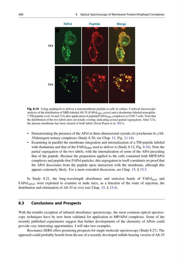
• Examining in parallel the membrane integration and internalization of a TM peptide labeled
with rhodamine and that of the FAPol NBD used to deliver it (Study 8.12; Fig. 8.10). Note the
partial segregation of the two labels, with the internalization of some of the APol preceding
that of the peptide. Because the preparation applied to the cells contained both MP/FAPol
complexes and peptide-free FAPol particles, this segregation in itself constitutes no proof that
the APol dissociates from the peptide upon interaction with the membrane, although this
appears extremely likely. For a more extended discussion, see Chap. 15, § 15.3.
In Study 8.21, the long-wavelength absorbance and emission bands of FAPol rhod and
FAPol AF647 were exploited to examine in nude mice, as a function of the route of injection, the
distribution and elimination of A8-35 in vivo (see Chap. 15, § 15.4).
8.3
Conclusions and Prospects
With the notable exception of infrared absorbance spectroscopy, the most common optical spectroscopy techniques have by now been validated for application to MP/APol complexes. Some of the
recently published experiments suggest that further developments of the chemistry of APols could
provide very interesting opportunities. I will take two examples.
Resonance SERS offers promising prospects for single-molecule spectroscopy (Study 8.27). The
approach could probably benefit from the use of a recently developed sulfide-bearing version of A8-35
Fig. 8.10 Using amphipols to deliver a transmembrane peptide to cells in culture. Confocal microscopic
analysis of the distribution of NBD-labeled A8-35 (FAPol NBD ; green) and a rhodamine-labeled neuropilin1 TM peptide (red) 16 and 72 h after application of peptide/FAPol NBD complexes to COS-7 cells. Note that
the distribution of the two labels does not totally overlap, indicating at least partial segregation. After 72 h,
the plasma membrane has been cleared of both labels (From Popot et al. 2011).
400
8 Optical Spectroscopy of Membrane Protein/Amphipol Complexes
• Examining in parallel the membrane integration and internalization of a TM peptide labeled
with rhodamine and that of the FAPol NBD used to deliver it (Study 8.12; Fig. 8.10). Note the
partial segregation of the two labels, with the internalization of some of the APol preceding
that of the peptide. Because the preparation applied to the cells contained both MP/FAPol
complexes and peptide-free FAPol particles, this segregation in itself constitutes no proof that
the APol dissociates from the peptide upon interaction with the membrane, although this
appears extremely likely. For a more extended discussion, see Chap. 15, § 15.3.
In Study 8.21, the long-wavelength absorbance and emission bands of FAPol rhod and
FAPol AF647 were exploited to examine in nude mice, as a function of the route of injection, the
distribution and elimination of A8-35 in vivo (see Chap. 15, § 15.4).
8.3
Conclusions and Prospects
With the notable exception of infrared absorbance spectroscopy, the most common optical spectroscopy techniques have by now been validated for application to MP/APol complexes. Some of the
recently published experiments suggest that further developments of the chemistry of APols could
provide very interesting opportunities. I will take two examples.
Resonance SERS offers promising prospects for single-molecule spectroscopy (Study 8.27). The
approach could probably benefit from the use of a recently developed sulfide-bearing version of A8-35
Fig. 8.10 Using amphipols to deliver a transmembrane peptide to cells in culture. Confocal microscopic
analysis of the distribution of NBD-labeled A8-35 (FAPol NBD ; green) and a rhodamine-labeled neuropilin1 TM peptide (red) 16 and 72 h after application of peptide/FAPol NBD complexes to COS-7 cells. Note that
the distribution of the two labels does not totally overlap, indicating at least partial segregation. After 72 h,
the plasma membrane has been cleared of both labels (From Popot et al. 2011).
400
8 Optical Spectroscopy of Membrane Protein/Amphipol Complexes