

8.2.2
Infrared and Raman Spectroscopy
No infrared absorbance spectroscopy data on APol-trapped MPs has been reported yet. The reason is
that the most widely used APols, including A8-35, all feature amide bonds (see Chap. 4). Their
vibration frequencies superimpose those of the peptide bonds, whose relative frequencies and
intensities are the main source of information that IR spectroscopy contributes to exploring the
structure of proteins. In principle, it is possible to distinguish between protein and APol amide
bonds by labeling one or the other partner with
15
N, which slightly changes some of the vibrational
frequencies of the amide bonds. This approach has been attempted by Yann Gohon and Erik
Goormaghtigh, using an
15 N-labeled MP trapped by unlabeled A8-35, but it has not succeeded in
sufficiently resolving the contributions of the two partners. Indeed, in a BR/A8-35 complex, (i) there is
~2 g A8-35 per g BR (Chap. 5) and (ii) for each kDa of material, a protein contributes ~9.09 and
A8-35 ~5.25 amide bonds. The ratio of the two contributions – assuming the complexes to have been
totally freed of unbound A8-35 – would therefore be ~0.87:1. Isotopic labeling does not separate
efficiently enough the bands contributed by the two partners to permit a clean resolution of the
protein’s bands. N-methylated A8-35 has recently been synthesized (F. Giusti, unpublished results),
which could perhaps solve this problem.
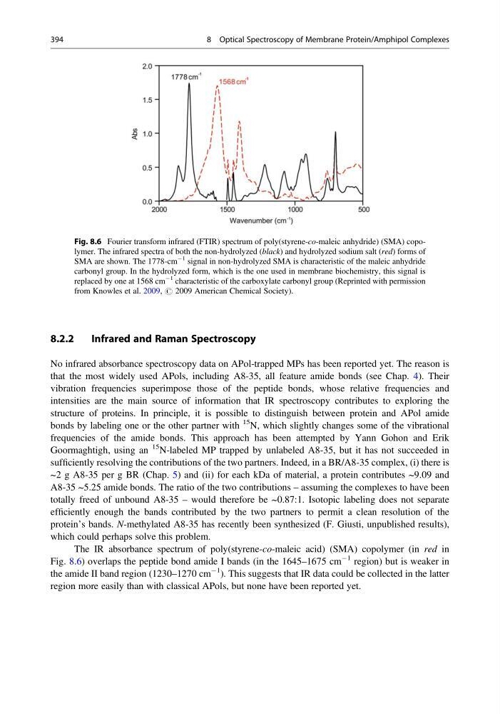
The IR absorbance spectrum of poly(styrene-co-maleic acid) (SMA) copolymer (in red in
Fig. 8.6) overlaps the peptide bond amide I bands (in the 1645–1675 cm
À1 region) but is weaker in
the amide II band region (1230–1270 cm
À1 ). This suggests that IR data could be collected in the latter
region more easily than with classical APols, but none have been reported yet.
Fig. 8.6 Fourier transform infrared (FTIR) spectrum of poly(styrene-co-maleic anhydride) (SMA) copolymer. The infrared spectra of both the non-hydrolyzed (black) and hydrolyzed sodium salt (red) forms of
SMA are shown. The 1778-cm
À1 signal in non-hydrolyzed SMA is characteristic of the maleic anhydride
carbonyl group. In the hydrolyzed form, which is the one used in membrane biochemistry, this signal is
replaced by one at 1568 cm
À1 characteristic of the carboxylate carbonyl group (Reprinted with permission
from Knowles et al. 2009, # 2009 American Chemical Society).
394
8 Optical Spectroscopy of Membrane Protein/Amphipol Complexes
Infrared and Raman Spectroscopy
No infrared absorbance spectroscopy data on APol-trapped MPs has been reported yet. The reason is
that the most widely used APols, including A8-35, all feature amide bonds (see Chap. 4). Their
vibration frequencies superimpose those of the peptide bonds, whose relative frequencies and
intensities are the main source of information that IR spectroscopy contributes to exploring the
structure of proteins. In principle, it is possible to distinguish between protein and APol amide
bonds by labeling one or the other partner with
15
N, which slightly changes some of the vibrational
frequencies of the amide bonds. This approach has been attempted by Yann Gohon and Erik
Goormaghtigh, using an
15 N-labeled MP trapped by unlabeled A8-35, but it has not succeeded in
sufficiently resolving the contributions of the two partners. Indeed, in a BR/A8-35 complex, (i) there is
~2 g A8-35 per g BR (Chap. 5) and (ii) for each kDa of material, a protein contributes ~9.09 and
A8-35 ~5.25 amide bonds. The ratio of the two contributions – assuming the complexes to have been
totally freed of unbound A8-35 – would therefore be ~0.87:1. Isotopic labeling does not separate
efficiently enough the bands contributed by the two partners to permit a clean resolution of the
protein’s bands. N-methylated A8-35 has recently been synthesized (F. Giusti, unpublished results),
which could perhaps solve this problem.
The IR absorbance spectrum of poly(styrene-co-maleic acid) (SMA) copolymer (in red in
Fig. 8.6) overlaps the peptide bond amide I bands (in the 1645–1675 cm
À1 region) but is weaker in
the amide II band region (1230–1270 cm
À1 ). This suggests that IR data could be collected in the latter
region more easily than with classical APols, but none have been reported yet.
Fig. 8.6 Fourier transform infrared (FTIR) spectrum of poly(styrene-co-maleic anhydride) (SMA) copolymer. The infrared spectra of both the non-hydrolyzed (black) and hydrolyzed sodium salt (red) forms of
SMA are shown. The 1778-cm
À1 signal in non-hydrolyzed SMA is characteristic of the maleic anhydride
carbonyl group. In the hydrolyzed form, which is the one used in membrane biochemistry, this signal is
replaced by one at 1568 cm
À1 characteristic of the carboxylate carbonyl group (Reprinted with permission
from Knowles et al. 2009, # 2009 American Chemical Society).
394
8 Optical Spectroscopy of Membrane Protein/Amphipol Complexes