

Another classical application of CD is to follow as a function of time and temperature the
denaturation of an APol-trapped vs. a detergent-solubilized or a membrane-embedded MP. This is
illustrated in Fig. 8.4 in the case of MOMP, the major outer membrane protein from the pathogenic
bacterium Chlamydia trachomatis (Study 8.11).
Visible CD spectra have been exploited to explore interactions between chromophores in
BR/SMALP complexes (Study 8.18; Fig. 8.5) and in APol-trapped light-harvesting complex II
(LHCII; Study 8.25). The bilobed CD spectrum of BR in purple membranes is generally attributed
to excitonic interactions between retinal molecules in the trimers that, associated with lipids, organize
into two-dimensional BR crystals to form purple membrane patches. This feature disappears in
detergent-solubilized BR (Heyn et al. 1975) and in SMALP-trapped BR, or BR reconstituted into
lipid bilayers (Fig. 8.5), consistent with the monomerization of BR.
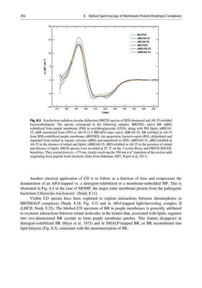
Fig. 8.3 Synchrotron radiation circular dichroism (SRCD) spectra of SDS-denatured and A8-35-refolded
bacteriorhodopsin. The spectra correspond to the following samples: BR/OTG, native BR (nBR)
solubilized from purple membrane (PM) in octylthioglucoside (OTG), along with PM lipids; nBR/A835, nBR transferred from OTG to A8-35 (1:5 BR/APol mass ratio); rBR/A8-35, BR refolded in A8-35
from SDS-solubilized purple membrane; dBO/SDS, the apoprotein, bacterio-opsin (BO), delipidated and
separated from retinal in organic solvents (dBO) and transferred to SDS; rdBO/A8-35, dBO refolded in
A8-35 in the absence of retinal and lipids; rdBR/A8-35, dBO refolded in A8-35 in the presence of retinal
and absence of lipids. SRCD spectra were recorded at 25
C on the 3 m-nim Bessy and DISCO SOLEIL
beamlines. They extend down to ~175 nm, clearly resolving the 190-nm π-π* transition of the exciton split
originating from peptide bond electrons (Data from Dahmane 2007, Popot et al. 2011).
392
8 Optical Spectroscopy of Membrane Protein/Amphipol Complexes
denaturation of an APol-trapped vs. a detergent-solubilized or a membrane-embedded MP. This is
illustrated in Fig. 8.4 in the case of MOMP, the major outer membrane protein from the pathogenic
bacterium Chlamydia trachomatis (Study 8.11).
Visible CD spectra have been exploited to explore interactions between chromophores in
BR/SMALP complexes (Study 8.18; Fig. 8.5) and in APol-trapped light-harvesting complex II
(LHCII; Study 8.25). The bilobed CD spectrum of BR in purple membranes is generally attributed
to excitonic interactions between retinal molecules in the trimers that, associated with lipids, organize
into two-dimensional BR crystals to form purple membrane patches. This feature disappears in
detergent-solubilized BR (Heyn et al. 1975) and in SMALP-trapped BR, or BR reconstituted into
lipid bilayers (Fig. 8.5), consistent with the monomerization of BR.
Fig. 8.3 Synchrotron radiation circular dichroism (SRCD) spectra of SDS-denatured and A8-35-refolded
bacteriorhodopsin. The spectra correspond to the following samples: BR/OTG, native BR (nBR)
solubilized from purple membrane (PM) in octylthioglucoside (OTG), along with PM lipids; nBR/A835, nBR transferred from OTG to A8-35 (1:5 BR/APol mass ratio); rBR/A8-35, BR refolded in A8-35
from SDS-solubilized purple membrane; dBO/SDS, the apoprotein, bacterio-opsin (BO), delipidated and
separated from retinal in organic solvents (dBO) and transferred to SDS; rdBO/A8-35, dBO refolded in
A8-35 in the absence of retinal and lipids; rdBR/A8-35, dBO refolded in A8-35 in the presence of retinal
and absence of lipids. SRCD spectra were recorded at 25
C on the 3 m-nim Bessy and DISCO SOLEIL
beamlines. They extend down to ~175 nm, clearly resolving the 190-nm π-π* transition of the exciton split
originating from peptide bond electrons (Data from Dahmane 2007, Popot et al. 2011).
392
8 Optical Spectroscopy of Membrane Protein/Amphipol Complexes