

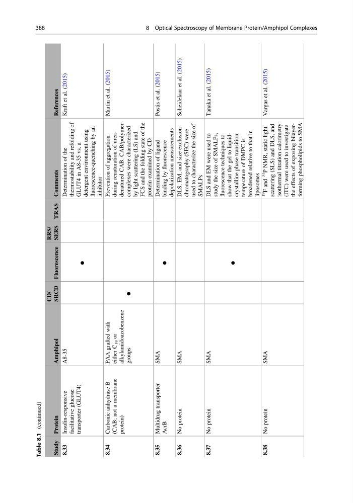
Table 8.1
(continued)
Study Protein
Amphipol
CD/
SRCD Fluorescence
RRS/
SERS TRAS Comments
References
8.33
Insulin-responsive
facilitative glucose
transporter (GLUT4)
A8-35
●
Determination of the
thermostability and refolding of
GLUT4 in A8-35 vs. a
detergent environment using
fluorescence-quenching by an
inhibitor
Kraft et al. (2015)
8.34
Carbonic anhydrase B
(CAB; not a membrane
protein)
PAA grafted with
either C
18 or
alkylamidoazobenzene
groups
●
Prevention of aggregation
during renaturation of ureadenatured CAB. CAB/polymer
complexes were characterized
by light scattering (LS) and
FCS and the folding state of the
protein examined by CD
Martin et al. (2015)
8.35
Multidrug transporter
AcrB
SMA
●
Determination of ligand
binding by
fluorescence
depolarization measurements
Postis et al. (2015)
8.36
No protein
SMA
DLS, EM, and size exclusion
chromatography (SEC) were
used to characterize the size of
SMALPs
Scheidelaar et al. (2015)
8.37
No protein
SMA
●
DLS and EM were used to
study the size of SMALPs,
fluorescence techniques to
show that the gel to liquidcrystalline phase transition
temperature of DMPC is
broadened relative to that in
liposomes
Tanaka et al. (2015)
8.38
No protein
SMA
19
F and
31
P NMR, static light
scattering (SLS) and DLS, and
isothermal titration calorimetry
(ITC) were used to investigate
the effects of exposing bilayerforming phospholipids to SMA
Vargas et al. (2015)
388
8 Optical Spectroscopy of Membrane Protein/Amphipol Complexes
(continued)
Study Protein
Amphipol
CD/
SRCD Fluorescence
RRS/
SERS TRAS Comments
References
8.33
Insulin-responsive
facilitative glucose
transporter (GLUT4)
A8-35
●
Determination of the
thermostability and refolding of
GLUT4 in A8-35 vs. a
detergent environment using
fluorescence-quenching by an
inhibitor
Kraft et al. (2015)
8.34
Carbonic anhydrase B
(CAB; not a membrane
protein)
PAA grafted with
either C
18 or
alkylamidoazobenzene
groups
●
Prevention of aggregation
during renaturation of ureadenatured CAB. CAB/polymer
complexes were characterized
by light scattering (LS) and
FCS and the folding state of the
protein examined by CD
Martin et al. (2015)
8.35
Multidrug transporter
AcrB
SMA
●
Determination of ligand
binding by
fluorescence
depolarization measurements
Postis et al. (2015)
8.36
No protein
SMA
DLS, EM, and size exclusion
chromatography (SEC) were
used to characterize the size of
SMALPs
Scheidelaar et al. (2015)
8.37
No protein
SMA
●
DLS and EM were used to
study the size of SMALPs,
fluorescence techniques to
show that the gel to liquidcrystalline phase transition
temperature of DMPC is
broadened relative to that in
liposomes
Tanaka et al. (2015)
8.38
No protein
SMA
19
F and
31
P NMR, static light
scattering (SLS) and DLS, and
isothermal titration calorimetry
(ITC) were used to investigate
the effects of exposing bilayerforming phospholipids to SMA
Vargas et al. (2015)
388
8 Optical Spectroscopy of Membrane Protein/Amphipol Complexes