

Nevertheless, the diversity of three-dimensional organizations that has now been revealed makes it
possible to propose some general rules.
Before moving on to the TM organization of MPs and their interactions between themselves and
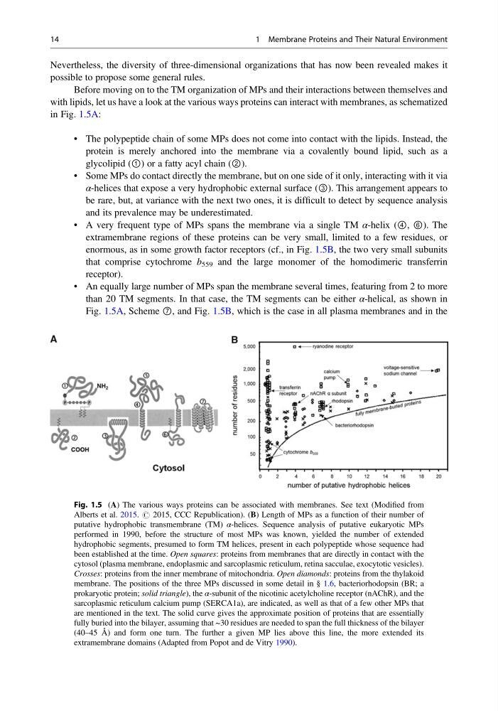
with lipids, let us have a look at the various ways proteins can interact with membranes, as schematized
in Fig. 1.5A:
• The polypeptide chain of some MPs does not come into contact with the lipids. Instead, the
protein is merely anchored into the membrane via a covalently bound lipid, such as a
glycolipid (①) or a fatty acyl chain (②).
• Some MPs do contact directly the membrane, but on one side of it only, interacting with it via
α-helices that expose a very hydrophobic external surface (③). This arrangement appears to
be rare, but, at variance with the next two ones, it is difficult to detect by sequence analysis
and its prevalence may be underestimated.
• A very frequent type of MPs spans the membrane via a single TM α-helix (④, ⑥). The
extramembrane regions of these proteins can be very small, limited to a few residues, or
enormous, as in some growth factor receptors (cf., in Fig. 1.5B, the two very small subunits
that comprise cytochrome b 559 and the large monomer of the homodimeric transferrin
receptor).
• An equally large number of MPs span the membrane several times, featuring from 2 to more
than 20 TM segments. In that case, the TM segments can be either α-helical, as shown in
Fig. 1.5A, Scheme ⑦, and Fig. 1.5B, which is the case in all plasma membranes and in the
Fig. 1.5 (A) The various ways proteins can be associated with membranes. See text (Modified from
Alberts et al. 2015. # 2015, CCC Republication). (B) Length of MPs as a function of their number of
putative hydrophobic transmembrane (TM) α-helices. Sequence analysis of putative eukaryotic MPs
performed in 1990, before the structure of most MPs was known, yielded the number of extended
hydrophobic segments, presumed to form TM helices, present in each polypeptide whose sequence had
been established at the time. Open squares: proteins from membranes that are directly in contact with the
cytosol (plasma membrane, endoplasmic and sarcoplasmic reticulum, retina sacculae, exocytotic vesicles).
Crosses: proteins from the inner membrane of mitochondria. Open diamonds: proteins from the thylakoid
membrane. The positions of the three MPs discussed in some detail in § 1.6, bacteriorhodopsin (BR; a
prokaryotic protein; solid triangle), the α-subunit of the nicotinic acetylcholine receptor (nAChR), and the
sarcoplasmic reticulum calcium pump (SERCA1a), are indicated, as well as that of a few other MPs that
are mentioned in the text. The solid curve gives the approximate position of proteins that are essentially
fully buried into the bilayer, assuming that ~30 residues are needed to span the full thickness of the bilayer
(40–45 Å) and form one turn. The further a given MP lies above this line, the more extended its
extramembrane domains (Adapted from Popot and de Vitry 1990).
14
1 Membrane Proteins and Their Natural Environment
possible to propose some general rules.
Before moving on to the TM organization of MPs and their interactions between themselves and
with lipids, let us have a look at the various ways proteins can interact with membranes, as schematized
in Fig. 1.5A:
• The polypeptide chain of some MPs does not come into contact with the lipids. Instead, the
protein is merely anchored into the membrane via a covalently bound lipid, such as a
glycolipid (①) or a fatty acyl chain (②).
• Some MPs do contact directly the membrane, but on one side of it only, interacting with it via
α-helices that expose a very hydrophobic external surface (③). This arrangement appears to
be rare, but, at variance with the next two ones, it is difficult to detect by sequence analysis
and its prevalence may be underestimated.
• A very frequent type of MPs spans the membrane via a single TM α-helix (④, ⑥). The
extramembrane regions of these proteins can be very small, limited to a few residues, or
enormous, as in some growth factor receptors (cf., in Fig. 1.5B, the two very small subunits
that comprise cytochrome b 559 and the large monomer of the homodimeric transferrin
receptor).
• An equally large number of MPs span the membrane several times, featuring from 2 to more
than 20 TM segments. In that case, the TM segments can be either α-helical, as shown in
Fig. 1.5A, Scheme ⑦, and Fig. 1.5B, which is the case in all plasma membranes and in the
Fig. 1.5 (A) The various ways proteins can be associated with membranes. See text (Modified from
Alberts et al. 2015. # 2015, CCC Republication). (B) Length of MPs as a function of their number of
putative hydrophobic transmembrane (TM) α-helices. Sequence analysis of putative eukaryotic MPs
performed in 1990, before the structure of most MPs was known, yielded the number of extended
hydrophobic segments, presumed to form TM helices, present in each polypeptide whose sequence had
been established at the time. Open squares: proteins from membranes that are directly in contact with the
cytosol (plasma membrane, endoplasmic and sarcoplasmic reticulum, retina sacculae, exocytotic vesicles).
Crosses: proteins from the inner membrane of mitochondria. Open diamonds: proteins from the thylakoid
membrane. The positions of the three MPs discussed in some detail in § 1.6, bacteriorhodopsin (BR; a
prokaryotic protein; solid triangle), the α-subunit of the nicotinic acetylcholine receptor (nAChR), and the
sarcoplasmic reticulum calcium pump (SERCA1a), are indicated, as well as that of a few other MPs that
are mentioned in the text. The solid curve gives the approximate position of proteins that are essentially
fully buried into the bilayer, assuming that ~30 residues are needed to span the full thickness of the bilayer
(40–45 Å) and form one turn. The further a given MP lies above this line, the more extended its
extramembrane domains (Adapted from Popot and de Vitry 1990).
14
1 Membrane Proteins and Their Natural Environment