

• All sorts of MPs have been shown to be amenable to folding in vitro: prokaryotic or
eukaryotic, α-helical bundles or β-barrels, monomeric or oligomeric, comprising or devoid
of prosthetic groups, etc. (Fig. 6.2).
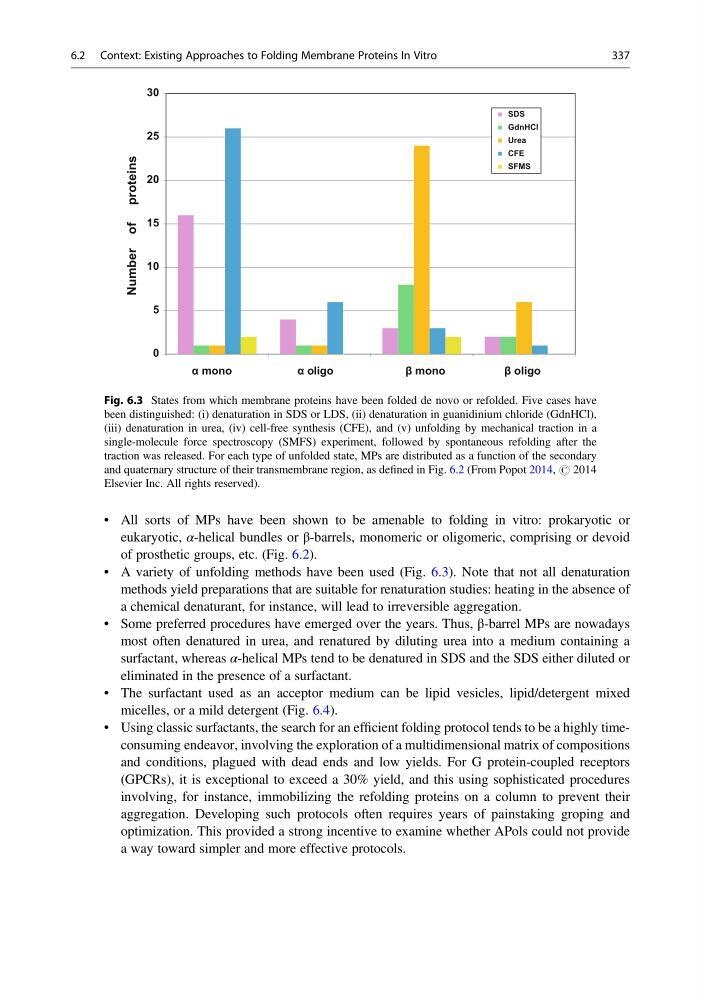
• A variety of unfolding methods have been used (Fig. 6.3). Note that not all denaturation
methods yield preparations that are suitable for renaturation studies: heating in the absence of
a chemical denaturant, for instance, will lead to irreversible aggregation.
• Some preferred procedures have emerged over the years. Thus, β-barrel MPs are nowadays
most often denatured in urea, and renatured by diluting urea into a medium containing a
surfactant, whereas α-helical MPs tend to be denatured in SDS and the SDS either diluted or
eliminated in the presence of a surfactant.
• The surfactant used as an acceptor medium can be lipid vesicles, lipid/detergent mixed
micelles, or a mild detergent (Fig. 6.4).
• Using classic surfactants, the search for an efficient folding protocol tends to be a highly timeconsuming endeavor, involving the exploration of a multidimensional matrix of compositions
and conditions, plagued with dead ends and low yields. For G protein-coupled receptors
(GPCRs), it is exceptional to exceed a 30% yield, and this using sophisticated procedures
involving, for instance, immobilizing the refolding proteins on a column to prevent their
aggregation. Developing such protocols often requires years of painstaking groping and
optimization. This provided a strong incentive to examine whether APols could not provide
a way toward simpler and more effective protocols.
0
5
10
15
20
25
30
SDS
GdnHCl
Urea
CFE
SFMS
Number of proteins
α mono
α oligo
β mono
β oligo
Fig. 6.3 States from which membrane proteins have been folded de novo or refolded. Five cases have
been distinguished: (i) denaturation in SDS or LDS, (ii) denaturation in guanidinium chloride (GdnHCl),
(iii) denaturation in urea, (iv) cell-free synthesis (CFE), and (v) unfolding by mechanical traction in a
single-molecule force spectroscopy (SMFS) experiment, followed by spontaneous refolding after the
traction was released. For each type of unfolded state, MPs are distributed as a function of the secondary
and quaternary structure of their transmembrane region, as defined in Fig. 6.2 (From Popot 2014, # 2014
Elsevier Inc. All rights reserved).
6.2 Context: Existing Approaches to Folding Membrane Proteins In Vitro
337
eukaryotic, α-helical bundles or β-barrels, monomeric or oligomeric, comprising or devoid
of prosthetic groups, etc. (Fig. 6.2).
• A variety of unfolding methods have been used (Fig. 6.3). Note that not all denaturation
methods yield preparations that are suitable for renaturation studies: heating in the absence of
a chemical denaturant, for instance, will lead to irreversible aggregation.
• Some preferred procedures have emerged over the years. Thus, β-barrel MPs are nowadays
most often denatured in urea, and renatured by diluting urea into a medium containing a
surfactant, whereas α-helical MPs tend to be denatured in SDS and the SDS either diluted or
eliminated in the presence of a surfactant.
• The surfactant used as an acceptor medium can be lipid vesicles, lipid/detergent mixed
micelles, or a mild detergent (Fig. 6.4).
• Using classic surfactants, the search for an efficient folding protocol tends to be a highly timeconsuming endeavor, involving the exploration of a multidimensional matrix of compositions
and conditions, plagued with dead ends and low yields. For G protein-coupled receptors
(GPCRs), it is exceptional to exceed a 30% yield, and this using sophisticated procedures
involving, for instance, immobilizing the refolding proteins on a column to prevent their
aggregation. Developing such protocols often requires years of painstaking groping and
optimization. This provided a strong incentive to examine whether APols could not provide
a way toward simpler and more effective protocols.
0
5
10
15
20
25
30
SDS
GdnHCl
Urea
CFE
SFMS
Number of proteins
α mono
α oligo
β mono
β oligo
Fig. 6.3 States from which membrane proteins have been folded de novo or refolded. Five cases have
been distinguished: (i) denaturation in SDS or LDS, (ii) denaturation in guanidinium chloride (GdnHCl),
(iii) denaturation in urea, (iv) cell-free synthesis (CFE), and (v) unfolding by mechanical traction in a
single-molecule force spectroscopy (SMFS) experiment, followed by spontaneous refolding after the
traction was released. For each type of unfolded state, MPs are distributed as a function of the secondary
and quaternary structure of their transmembrane region, as defined in Fig. 6.2 (From Popot 2014, # 2014
Elsevier Inc. All rights reserved).
6.2 Context: Existing Approaches to Folding Membrane Proteins In Vitro
337