

α-helical structure (~50% vs. ~75% in the native protein; for a recent review on MP/SDS complexes, see
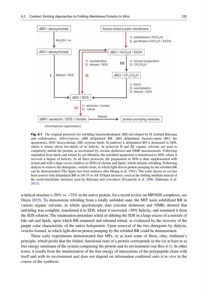
Otzen 2015). To demonstrate refolding from a totally unfolded state, the MIT team solubilized BR in
various organic solvents, in which spectroscopic data (circular dichroism and NMR) showed that
unfolding was complete, transferred it to SDS, where it recovered ~50% helicity, and renatured it from
the SDS solution. The renaturation procedure relied on diluting the SDS in a large excess of a mixture of
bile salt and lipids, upon which BR renatured and rebound retinal, as evidenced by the recovery of the
purple color characteristic of the native holoprotein. Upon removal of the two detergents by dialysis,
vesicles formed, in which light-driven proton pumping by the refolded BR could be demonstrated.
These early experiments demonstrated that MPs, or at least some of them, obey Anfinsen’s
principle, which posits that the folded, functional state of a protein corresponds to the (or at least to a)
free energy minimum of the system comprising the protein and its environment (see Box 6.1). In other
terms, it results from the minimization of the free energy of interactions of the polypeptide chain with
itself and with its environment and does not depend on information conferred onto it in vivo in the
course of the synthesis.
Fig. 6.1 The original protocols for refolding bacteriorhodopsin (BR) developed by H. Gobind Khorana
and collaborators. Abbreviations: dBR delipidated BR, dBO delipidated bacterio-opsin (BO; the
apoprotein), DOC deoxycholate, SBL soybean lipids. In pathway I, delipidated BO is denatured in SDS,
where it retains about two-thirds of its helicity. In protocols II and III, organic solvents are used to
completely unfold the protein, as ascertained by circular dichroism and NMR measurements. Following
separation from lipids and retinal by gel filtration, the unfolded apoprotein is transferred to SDS, where it
recovers a degree of helicity. In all three protocols, the preparation in SDS is then supplemented with
retinal and with a large excess (relative to SDS) of cholate and lipids, which initiates refolding. Following
dialysis to remove the detergents, vesicles form, in which light-driven proton pumping by the refolded BR
can be demonstrated (The figure has been redrawn after Huang et al. 1981). The route shown in red has
been used to fold delipidated BR in A8-35 or A8-35/lipid mixtures, used as the folding medium instead of
the asolectin/cholate mixtures used by Khorana and coworkers (Pocanschi et al. 2006; Dahmane et al.
2013).
6.2 Context: Existing Approaches to Folding Membrane Proteins In Vitro
335
Otzen 2015). To demonstrate refolding from a totally unfolded state, the MIT team solubilized BR in
various organic solvents, in which spectroscopic data (circular dichroism and NMR) showed that
unfolding was complete, transferred it to SDS, where it recovered ~50% helicity, and renatured it from
the SDS solution. The renaturation procedure relied on diluting the SDS in a large excess of a mixture of
bile salt and lipids, upon which BR renatured and rebound retinal, as evidenced by the recovery of the
purple color characteristic of the native holoprotein. Upon removal of the two detergents by dialysis,
vesicles formed, in which light-driven proton pumping by the refolded BR could be demonstrated.
These early experiments demonstrated that MPs, or at least some of them, obey Anfinsen’s
principle, which posits that the folded, functional state of a protein corresponds to the (or at least to a)
free energy minimum of the system comprising the protein and its environment (see Box 6.1). In other
terms, it results from the minimization of the free energy of interactions of the polypeptide chain with
itself and with its environment and does not depend on information conferred onto it in vivo in the
course of the synthesis.
Fig. 6.1 The original protocols for refolding bacteriorhodopsin (BR) developed by H. Gobind Khorana
and collaborators. Abbreviations: dBR delipidated BR, dBO delipidated bacterio-opsin (BO; the
apoprotein), DOC deoxycholate, SBL soybean lipids. In pathway I, delipidated BO is denatured in SDS,
where it retains about two-thirds of its helicity. In protocols II and III, organic solvents are used to
completely unfold the protein, as ascertained by circular dichroism and NMR measurements. Following
separation from lipids and retinal by gel filtration, the unfolded apoprotein is transferred to SDS, where it
recovers a degree of helicity. In all three protocols, the preparation in SDS is then supplemented with
retinal and with a large excess (relative to SDS) of cholate and lipids, which initiates refolding. Following
dialysis to remove the detergents, vesicles form, in which light-driven proton pumping by the refolded BR
can be demonstrated (The figure has been redrawn after Huang et al. 1981). The route shown in red has
been used to fold delipidated BR in A8-35 or A8-35/lipid mixtures, used as the folding medium instead of
the asolectin/cholate mixtures used by Khorana and coworkers (Pocanschi et al. 2006; Dahmane et al.
2013).
6.2 Context: Existing Approaches to Folding Membrane Proteins In Vitro
335