

interior, the matrix, is packed with stacks of sacculae in which the transduction of light into chemical
energy takes place, thanks to oxygenic photosynthesis.
Each of the membranes that compartmentalize a eukaryotic cell has its own lipid and protein
composition, and its internal fluid also has a specific composition. For instance, the cytosol is a
reducing medium, whereas the lumen of the endoplasmic reticulum is, as the exterior medium,
oxidizing. The concentration of calcium ions is very low in the cytosol, very high in the lumen of
the sarcoplasmic reticulum (SR); the electrochemical potential of H 3 O
+ ions is higher in the lumen of
chloroplasts and in the intermembrane space of mitochondria than in their matrix, etc.
Biological membranes do not comprise a single type of lipid but mixtures of them, in which both
the polar head hydrophobic chains vary. The chains – most often two of them but sometimes more
(cf. Fig. 1.1) – can be long or short (typically between 16- and 22-carbon long), saturated or
unsaturated, sometimes branched (in archaebacteria). They can be associated to the polar head either
by hydrolyzable ester functions (in eubacteria and eukaryotes) or non-hydrolyzable ether functions
(in archaebacteria). The polar heads can be zwitterionic, as in PC or phosphatidylethanolamine (PE),
and nonionic (formed of sugar residues) or carry a net negative charge, as in phosphatidylserine (PS)
and phosphatidylglycerol (PG) (Fig. 1.1). In these glycerophospholipids, in bulk the most abundant
lipids in animal cells, two acyl chains and a polar head are attached, respectively, to the first two and to
the third one of the three hydroxyl functions of glycerol. In sphingomyelin (Sph), one acyl chain is
linked by an amide bond to a ceramide group, which is comprised of an alkyl chain and a polar head
(Fig. 1.1). Eukaryotic membranes also contain sterols, such as cholesterol, in animal cells, or
ergosterol, in yeasts (Fig. 1.1). By themselves, sterols do not form bilayers, but they partition into
them and modulate their thickness and fluidity. In a living cell, the composition of the lipids is adjusted
so that they remain in a fluid phase. This is achieved by modulating the length and level of unsaturation
of the chains so that they remain mobile with respect to one another and do not associate into a gel.
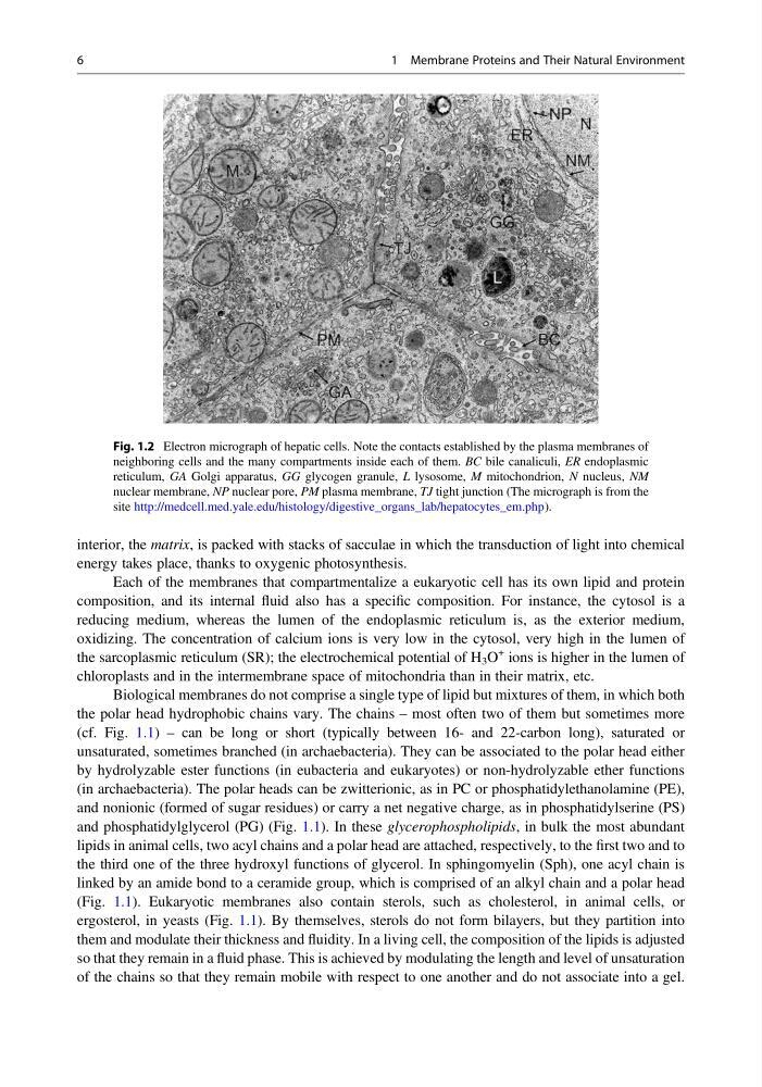
Fig. 1.2 Electron micrograph of hepatic cells. Note the contacts established by the plasma membranes of
neighboring cells and the many compartments inside each of them. BC bile canaliculi, ER endoplasmic
reticulum, GA Golgi apparatus, GG glycogen granule, L lysosome, M mitochondrion, N nucleus, NM
nuclear membrane, NP nuclear pore, PM plasma membrane, TJ tight junction (The micrograph is from the
site http://medcell.med.yale.edu/histology/digestive_organs_lab/hepatocytes_em.php).
6
1 Membrane Proteins and Their Natural Environment
energy takes place, thanks to oxygenic photosynthesis.
Each of the membranes that compartmentalize a eukaryotic cell has its own lipid and protein
composition, and its internal fluid also has a specific composition. For instance, the cytosol is a
reducing medium, whereas the lumen of the endoplasmic reticulum is, as the exterior medium,
oxidizing. The concentration of calcium ions is very low in the cytosol, very high in the lumen of
the sarcoplasmic reticulum (SR); the electrochemical potential of H 3 O
+ ions is higher in the lumen of
chloroplasts and in the intermembrane space of mitochondria than in their matrix, etc.
Biological membranes do not comprise a single type of lipid but mixtures of them, in which both
the polar head hydrophobic chains vary. The chains – most often two of them but sometimes more
(cf. Fig. 1.1) – can be long or short (typically between 16- and 22-carbon long), saturated or
unsaturated, sometimes branched (in archaebacteria). They can be associated to the polar head either
by hydrolyzable ester functions (in eubacteria and eukaryotes) or non-hydrolyzable ether functions
(in archaebacteria). The polar heads can be zwitterionic, as in PC or phosphatidylethanolamine (PE),
and nonionic (formed of sugar residues) or carry a net negative charge, as in phosphatidylserine (PS)
and phosphatidylglycerol (PG) (Fig. 1.1). In these glycerophospholipids, in bulk the most abundant
lipids in animal cells, two acyl chains and a polar head are attached, respectively, to the first two and to
the third one of the three hydroxyl functions of glycerol. In sphingomyelin (Sph), one acyl chain is
linked by an amide bond to a ceramide group, which is comprised of an alkyl chain and a polar head
(Fig. 1.1). Eukaryotic membranes also contain sterols, such as cholesterol, in animal cells, or
ergosterol, in yeasts (Fig. 1.1). By themselves, sterols do not form bilayers, but they partition into
them and modulate their thickness and fluidity. In a living cell, the composition of the lipids is adjusted
so that they remain in a fluid phase. This is achieved by modulating the length and level of unsaturation
of the chains so that they remain mobile with respect to one another and do not associate into a gel.
Fig. 1.2 Electron micrograph of hepatic cells. Note the contacts established by the plasma membranes of
neighboring cells and the many compartments inside each of them. BC bile canaliculi, ER endoplasmic
reticulum, GA Golgi apparatus, GG glycogen granule, L lysosome, M mitochondrion, N nucleus, NM
nuclear membrane, NP nuclear pore, PM plasma membrane, TJ tight junction (The micrograph is from the
site http://medcell.med.yale.edu/histology/digestive_organs_lab/hepatocytes_em.php).
6
1 Membrane Proteins and Their Natural Environment