

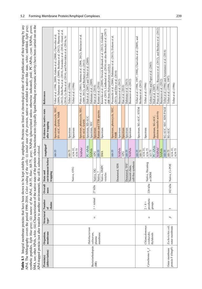
Table 5.1
Integral membrane proteins that have been shown to be kept soluble by amphipols. Proteins are listed in chronological order of first publication of their trapping by any
APol. The bibliography covers the period 1996–2016. Color coding of the cells:
(i) initial state of the protein,
native or refolded to native-like state, white;
unfolded, light gray;
synthetic peptides, light blue-gray;
(ii) nature of the APol,
A8-35, blue;
A8-75, gray;
NAPols (glucosylated unless otherwise indicated), pink;
PC-APols, cyan;
SAPols, green;
SMA, tan;
other APols, white.
(iii) Characterization of the native state of the protein: when functional tests (typically ligand binding or enzymatic activity) have been carried out on the
APol-trapped protein (not after transfer to another environment), the cell is salmon-colored.
Protein(s)
(abbreviation)
Organism,
membrane
Structural
type a
Number
of
chains
Overall
mass
State and
environment before
trapping
Amphipol b
Evidence for native state
after trapping
References
Bacteriorhodopsin
(BR)
Halobacterium
salinarum,
plasma
membrane
α
1 + retinal
27 kDa
Native, OTG
A8-35
Spectrum, photocycle, SEC,
SV-AUC, SANS, NMR
Tribet et al. (1996, 2009), Gohon et al. (2008), Charvolin et al.
(2009), Dahmane et al. (2009), Bechara et al. (2012), Sharma et al.
(2012), Della Pia et al. (2014), Ferrandez et al. (2014), Giusti et al.
(2014), Le Bon et al. (2014), and Polovinkin et al. (2014a, b)
A8-75
Spectrum
Tribet et al. (1996)
A34-35,
A34-75
Spectrum
Tribet et al. (1996)
NAPols c
Spectrum, photocycle, SEC,
SV-AUC, SANS
Prata et al. (2001), Sharma et al. (2008, 2012), Bazzacco et al.
(2009, 2012), and Bechara et al. (2012)
PC-APols
Spectrum, SD-AUC
Diab et al. (2007) and Tribet et al. (2009)
SAPols
Spectrum, SEC
Dahmane et al. (2009)
Native, OG
APG
Spectrum
Han et al. (2014)
Native, ?
A8-35
Raman and FTIR spectra
Kumar et al. (2016)
Native, DMPC
vesicles
SMA
Spectrum
Knowles et al. (2009), Orwick-Rydmark et al. (2012), Goddard
et al. (2015), Lindhoud et al. (2016) see also Broecker et al. (2017)
(BR from Haloquadratum walsbyi)
Denatured, SDS
A8-35
Spectrum, photocycle, SEC,
NMR
Pocanschi et al. (2006), Dahmane et al. (2013), Etzkorn et al.
(2013), and Elter et al. (2014)
NAPols
Spectrum
Bazzacco et al. (2012)
APG
Spectrum
Han et al. (2014)
Denatured, TFE
c
A8-35
Spectrum
Dahmane et al. (2013)
Nascent chain,
cell-free synthesis
NAPols
Spectrum
Bazzacco et al. (2012)
Cytochrome b
6
f
Chlamydomonas
reinhardtii,
thylakoids
α
2 × 8 +
prosthetic
groups
228 kDa
Native, Hecameg
or DDM
A8-35
Spectrum, SG-AUC, STEM
Tribet et al. (1996, 1997, 1998), Charvolin et al. (2009), and
Bechara et al. (2012)
A8-75
A34-35,
A34-75
Spectrum
Tribet et al. (1996)
A5-75
Spectrum, SG-AUC,
electron transfer activity
Gohon (1996) and Popot et al. (2003)
PC-APols
Diab et al. (2007)
NAPols
c
Prata et al. (2001), Bazzacco et al. (2012), and Bechara et al. (2012)
Bazzacco et al. (2012)
SAPols
Outer membrane
protein F (OmpF)
Escherichia coli,
outer membrane
β
3
102 kDa Native, C8-POE
A8-35
SG-AUC, SEC, SDS-PAGE Tribet et al. (1996) and Arunmanee et al. (2014)
A8-75
SG-AUC
Tribet et al. (1996, 1997)
A34-35,
A34-75
Tribet et al. (1996)
5.2 Forming Membrane Protein/Amphipol Complexes
239
Integral membrane proteins that have been shown to be kept soluble by amphipols. Proteins are listed in chronological order of first publication of their trapping by any
APol. The bibliography covers the period 1996–2016. Color coding of the cells:
(i) initial state of the protein,
native or refolded to native-like state, white;
unfolded, light gray;
synthetic peptides, light blue-gray;
(ii) nature of the APol,
A8-35, blue;
A8-75, gray;
NAPols (glucosylated unless otherwise indicated), pink;
PC-APols, cyan;
SAPols, green;
SMA, tan;
other APols, white.
(iii) Characterization of the native state of the protein: when functional tests (typically ligand binding or enzymatic activity) have been carried out on the
APol-trapped protein (not after transfer to another environment), the cell is salmon-colored.
Protein(s)
(abbreviation)
Organism,
membrane
Structural
type a
Number
of
chains
Overall
mass
State and
environment before
trapping
Amphipol b
Evidence for native state
after trapping
References
Bacteriorhodopsin
(BR)
Halobacterium
salinarum,
plasma
membrane
α
1 + retinal
27 kDa
Native, OTG
A8-35
Spectrum, photocycle, SEC,
SV-AUC, SANS, NMR
Tribet et al. (1996, 2009), Gohon et al. (2008), Charvolin et al.
(2009), Dahmane et al. (2009), Bechara et al. (2012), Sharma et al.
(2012), Della Pia et al. (2014), Ferrandez et al. (2014), Giusti et al.
(2014), Le Bon et al. (2014), and Polovinkin et al. (2014a, b)
A8-75
Spectrum
Tribet et al. (1996)
A34-35,
A34-75
Spectrum
Tribet et al. (1996)
NAPols c
Spectrum, photocycle, SEC,
SV-AUC, SANS
Prata et al. (2001), Sharma et al. (2008, 2012), Bazzacco et al.
(2009, 2012), and Bechara et al. (2012)
PC-APols
Spectrum, SD-AUC
Diab et al. (2007) and Tribet et al. (2009)
SAPols
Spectrum, SEC
Dahmane et al. (2009)
Native, OG
APG
Spectrum
Han et al. (2014)
Native, ?
A8-35
Raman and FTIR spectra
Kumar et al. (2016)
Native, DMPC
vesicles
SMA
Spectrum
Knowles et al. (2009), Orwick-Rydmark et al. (2012), Goddard
et al. (2015), Lindhoud et al. (2016) see also Broecker et al. (2017)
(BR from Haloquadratum walsbyi)
Denatured, SDS
A8-35
Spectrum, photocycle, SEC,
NMR
Pocanschi et al. (2006), Dahmane et al. (2013), Etzkorn et al.
(2013), and Elter et al. (2014)
NAPols
Spectrum
Bazzacco et al. (2012)
APG
Spectrum
Han et al. (2014)
Denatured, TFE
c
A8-35
Spectrum
Dahmane et al. (2013)
Nascent chain,
cell-free synthesis
NAPols
Spectrum
Bazzacco et al. (2012)
Cytochrome b
6
f
Chlamydomonas
reinhardtii,
thylakoids
α
2 × 8 +
prosthetic
groups
228 kDa
Native, Hecameg
or DDM
A8-35
Spectrum, SG-AUC, STEM
Tribet et al. (1996, 1997, 1998), Charvolin et al. (2009), and
Bechara et al. (2012)
A8-75
A34-35,
A34-75
Spectrum
Tribet et al. (1996)
A5-75
Spectrum, SG-AUC,
electron transfer activity
Gohon (1996) and Popot et al. (2003)
PC-APols
Diab et al. (2007)
NAPols
c
Prata et al. (2001), Bazzacco et al. (2012), and Bechara et al. (2012)
Bazzacco et al. (2012)
SAPols
Outer membrane
protein F (OmpF)
Escherichia coli,
outer membrane
β
3
102 kDa Native, C8-POE
A8-35
SG-AUC, SEC, SDS-PAGE Tribet et al. (1996) and Arunmanee et al. (2014)
A8-75
SG-AUC
Tribet et al. (1996, 1997)
A34-35,
A34-75
Tribet et al. (1996)
5.2 Forming Membrane Protein/Amphipol Complexes
239