

partners and the third one in distinct NDs (Shin et al. 2014). A related approach is to study the
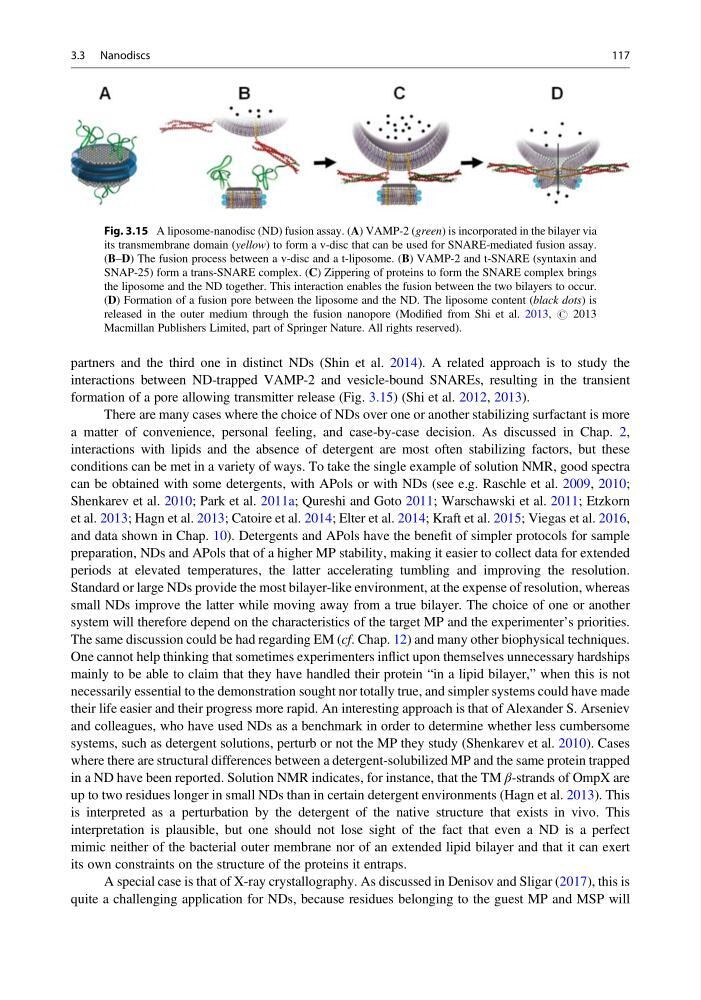
interactions between ND-trapped VAMP-2 and vesicle-bound SNAREs, resulting in the transient
formation of a pore allowing transmitter release (Fig. 3.15) (Shi et al. 2012, 2013).
There are many cases where the choice of NDs over one or another stabilizing surfactant is more
a matter of convenience, personal feeling, and case-by-case decision. As discussed in Chap. 2,
interactions with lipids and the absence of detergent are most often stabilizing factors, but these
conditions can be met in a variety of ways. To take the single example of solution NMR, good spectra
can be obtained with some detergents, with APols or with NDs (see e.g. Raschle et al. 2009, 2010;
Shenkarev et al. 2010; Park et al. 2011a; Qureshi and Goto 2011; Warschawski et al. 2011; Etzkorn
et al. 2013; Hagn et al. 2013; Catoire et al. 2014; Elter et al. 2014; Kraft et al. 2015; Viegas et al. 2016,
and data shown in Chap. 10). Detergents and APols have the benefit of simpler protocols for sample
preparation, NDs and APols that of a higher MP stability, making it easier to collect data for extended
periods at elevated temperatures, the latter accelerating tumbling and improving the resolution.
Standard or large NDs provide the most bilayer-like environment, at the expense of resolution, whereas
small NDs improve the latter while moving away from a true bilayer. The choice of one or another
system will therefore depend on the characteristics of the target MP and the experimenter’s priorities.
The same discussion could be had regarding EM (cf. Chap. 12) and many other biophysical techniques.
One cannot help thinking that sometimes experimenters inflict upon themselves unnecessary hardships
mainly to be able to claim that they have handled their protein “in a lipid bilayer,” when this is not
necessarily essential to the demonstration sought nor totally true, and simpler systems could have made
their life easier and their progress more rapid. An interesting approach is that of Alexander S. Arseniev
and colleagues, who have used NDs as a benchmark in order to determine whether less cumbersome
systems, such as detergent solutions, perturb or not the MP they study (Shenkarev et al. 2010). Cases
where there are structural differences between a detergent-solubilized MP and the same protein trapped
in a ND have been reported. Solution NMR indicates, for instance, that the TM β-strands of OmpX are
up to two residues longer in small NDs than in certain detergent environments (Hagn et al. 2013). This
is interpreted as a perturbation by the detergent of the native structure that exists in vivo. This
interpretation is plausible, but one should not lose sight of the fact that even a ND is a perfect
mimic neither of the bacterial outer membrane nor of an extended lipid bilayer and that it can exert
its own constraints on the structure of the proteins it entraps.
A special case is that of X-ray crystallography. As discussed in Denisov and Sligar (2017), this is
quite a challenging application for NDs, because residues belonging to the guest MP and MSP will
Fig. 3.15 A liposome-nanodisc (ND) fusion assay. (A) VAMP-2 (green) is incorporated in the bilayer via
its transmembrane domain (yellow) to form a v-disc that can be used for SNARE-mediated fusion assay.
(B–D) The fusion process between a v-disc and a t-liposome. (B) VAMP-2 and t-SNARE (syntaxin and
SNAP-25) form a trans-SNARE complex. (C) Zippering of proteins to form the SNARE complex brings
the liposome and the ND together. This interaction enables the fusion between the two bilayers to occur.
(D) Formation of a fusion pore between the liposome and the ND. The liposome content (black dots) is
released in the outer medium through the fusion nanopore (Modified from Shi et al. 2013, # 2013
Macmillan Publishers Limited, part of Springer Nature. All rights reserved).
3.3 Nanodiscs
117
interactions between ND-trapped VAMP-2 and vesicle-bound SNAREs, resulting in the transient
formation of a pore allowing transmitter release (Fig. 3.15) (Shi et al. 2012, 2013).
There are many cases where the choice of NDs over one or another stabilizing surfactant is more
a matter of convenience, personal feeling, and case-by-case decision. As discussed in Chap. 2,
interactions with lipids and the absence of detergent are most often stabilizing factors, but these
conditions can be met in a variety of ways. To take the single example of solution NMR, good spectra
can be obtained with some detergents, with APols or with NDs (see e.g. Raschle et al. 2009, 2010;
Shenkarev et al. 2010; Park et al. 2011a; Qureshi and Goto 2011; Warschawski et al. 2011; Etzkorn
et al. 2013; Hagn et al. 2013; Catoire et al. 2014; Elter et al. 2014; Kraft et al. 2015; Viegas et al. 2016,
and data shown in Chap. 10). Detergents and APols have the benefit of simpler protocols for sample
preparation, NDs and APols that of a higher MP stability, making it easier to collect data for extended
periods at elevated temperatures, the latter accelerating tumbling and improving the resolution.
Standard or large NDs provide the most bilayer-like environment, at the expense of resolution, whereas
small NDs improve the latter while moving away from a true bilayer. The choice of one or another
system will therefore depend on the characteristics of the target MP and the experimenter’s priorities.
The same discussion could be had regarding EM (cf. Chap. 12) and many other biophysical techniques.
One cannot help thinking that sometimes experimenters inflict upon themselves unnecessary hardships
mainly to be able to claim that they have handled their protein “in a lipid bilayer,” when this is not
necessarily essential to the demonstration sought nor totally true, and simpler systems could have made
their life easier and their progress more rapid. An interesting approach is that of Alexander S. Arseniev
and colleagues, who have used NDs as a benchmark in order to determine whether less cumbersome
systems, such as detergent solutions, perturb or not the MP they study (Shenkarev et al. 2010). Cases
where there are structural differences between a detergent-solubilized MP and the same protein trapped
in a ND have been reported. Solution NMR indicates, for instance, that the TM β-strands of OmpX are
up to two residues longer in small NDs than in certain detergent environments (Hagn et al. 2013). This
is interpreted as a perturbation by the detergent of the native structure that exists in vivo. This
interpretation is plausible, but one should not lose sight of the fact that even a ND is a perfect
mimic neither of the bacterial outer membrane nor of an extended lipid bilayer and that it can exert
its own constraints on the structure of the proteins it entraps.
A special case is that of X-ray crystallography. As discussed in Denisov and Sligar (2017), this is
quite a challenging application for NDs, because residues belonging to the guest MP and MSP will
Fig. 3.15 A liposome-nanodisc (ND) fusion assay. (A) VAMP-2 (green) is incorporated in the bilayer via
its transmembrane domain (yellow) to form a v-disc that can be used for SNARE-mediated fusion assay.
(B–D) The fusion process between a v-disc and a t-liposome. (B) VAMP-2 and t-SNARE (syntaxin and
SNAP-25) form a trans-SNARE complex. (C) Zippering of proteins to form the SNARE complex brings
the liposome and the ND together. This interaction enables the fusion between the two bilayers to occur.
(D) Formation of a fusion pore between the liposome and the ND. The liposome content (black dots) is
released in the outer medium through the fusion nanopore (Modified from Shi et al. 2013, # 2013
Macmillan Publishers Limited, part of Springer Nature. All rights reserved).
3.3 Nanodiscs
117