

Cell
Water
Evanescent
wave
Evanescent
wave
Evanescent image
Epifluorescence imgage
Glass
Reflected beam Incident beam
Prism
c
a
b
d
Mirror
Mirror
Focusing lens
Laser
Filter
Camera
Water phase
Epifluorescence
illumination
Data acquisition & processing
Filter
Hg lamp
Glass slide
Objective lens
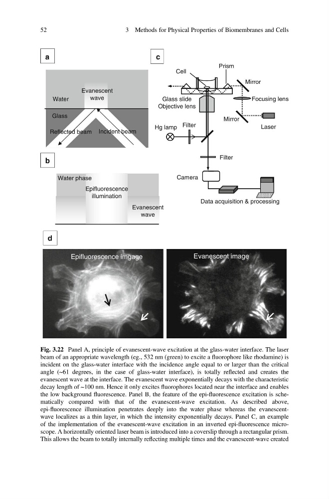
Fig. 3.22 Panel A, principle of evanescent-wave excitation at the glass-water interface. The laser
beam of an appropriate wavelength (eg., 532 nm (green) to excite a fluorophore like rhodamine) is
incident on the glass-water interface with the incidence angle equal to or larger than the critical
angle (~61 degrees, in the case of glass-water interface), is totally reflected and creates the
evanescent wave at the interface. The evanescent wave exponentially decays with the characteristic
decay length of ~100 nm. Hence it only excites fluorophores located near the interface and enables
the low background fluorescence. Panel B, the feature of the epi-fluorescence excitation is schematically compared with that of the evanescent-wave excitation. As described above,
epi-fluorescence illumination penetrates deeply into the water phase whereas the evanescentwave localizes as a thin layer, in which the intensity exponentially decays. Panel C, an example
of the implementation of the evanescent-wave excitation in an inverted epi-fluorescence microscope. A horizontally oriented laser beam is introduced into a coverslip through a rectangular prism.
This allows the beam to totally internally reflecting multiple times and the evanescent-wave created
52
3 Methods for Physical Properties of Biomembranes and Cells
Water
Evanescent
wave
Evanescent
wave
Evanescent image
Epifluorescence imgage
Glass
Reflected beam Incident beam
Prism
c
a
b
d
Mirror
Mirror
Focusing lens
Laser
Filter
Camera
Water phase
Epifluorescence
illumination
Data acquisition & processing
Filter
Hg lamp
Glass slide
Objective lens
Fig. 3.22 Panel A, principle of evanescent-wave excitation at the glass-water interface. The laser
beam of an appropriate wavelength (eg., 532 nm (green) to excite a fluorophore like rhodamine) is
incident on the glass-water interface with the incidence angle equal to or larger than the critical
angle (~61 degrees, in the case of glass-water interface), is totally reflected and creates the
evanescent wave at the interface. The evanescent wave exponentially decays with the characteristic
decay length of ~100 nm. Hence it only excites fluorophores located near the interface and enables
the low background fluorescence. Panel B, the feature of the epi-fluorescence excitation is schematically compared with that of the evanescent-wave excitation. As described above,
epi-fluorescence illumination penetrates deeply into the water phase whereas the evanescentwave localizes as a thin layer, in which the intensity exponentially decays. Panel C, an example
of the implementation of the evanescent-wave excitation in an inverted epi-fluorescence microscope. A horizontally oriented laser beam is introduced into a coverslip through a rectangular prism.
This allows the beam to totally internally reflecting multiple times and the evanescent-wave created
52
3 Methods for Physical Properties of Biomembranes and Cells