

References
1. F. Ritossa, Experimentia 18, 571 (1962)
2. R.N. McElhaney, K.A. Souza, Biochim. Biophys. Acta 443, 348 (1976)
3. H. Nakayama, M. Goto, K. Ohki, M. Mitsui, Y. Nozawa, Biochim. Biophys. Acta 730, 17 (1983)
4. H. Nakayama, K. Ohki, T. Mitsui, Y. Nozawa, Biochim. Biophys. Acta 769, 311 (1984)
5. M. Sinensky, Proc. Natl. Acad. Sci. USA 71, 522 (1974)
6. Y. Tasaka, Z. Gombos, Y. Nishiyama, T. Ohba, K. Ohki, N. Murata, EMBO J. 15, 641 (1996)
7. Y. Kameyama, K. Ohki, Y. Nozawa, J. Biochem. 88, 1291 (1980)
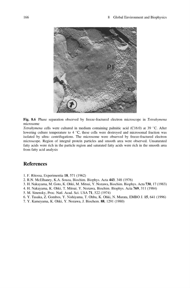
Fig. 8.6 Phase separation observed by freeze-fractured electron microscope in Tetrahymena
microsome
Tetrahymena cells were cultured in medium containing palmitic acid (C16:0) at 39
C. After
lowering culture temperature to 4
C, these cells were destroyed and microsomal fraction was
isolated by ultra- centrifugations. The microsome were observed by freeze-fractured electron
microscope. Region of integral protein particles and smooth area were observed. Unsaturated
fatty acids were rich in the particle region and saturated fatty acids were rich in the smooth area
from fatty acid analysis
166
8 Global Environment and Biophysics
1. F. Ritossa, Experimentia 18, 571 (1962)
2. R.N. McElhaney, K.A. Souza, Biochim. Biophys. Acta 443, 348 (1976)
3. H. Nakayama, M. Goto, K. Ohki, M. Mitsui, Y. Nozawa, Biochim. Biophys. Acta 730, 17 (1983)
4. H. Nakayama, K. Ohki, T. Mitsui, Y. Nozawa, Biochim. Biophys. Acta 769, 311 (1984)
5. M. Sinensky, Proc. Natl. Acad. Sci. USA 71, 522 (1974)
6. Y. Tasaka, Z. Gombos, Y. Nishiyama, T. Ohba, K. Ohki, N. Murata, EMBO J. 15, 641 (1996)
7. Y. Kameyama, K. Ohki, Y. Nozawa, J. Biochem. 88, 1291 (1980)
Fig. 8.6 Phase separation observed by freeze-fractured electron microscope in Tetrahymena
microsome
Tetrahymena cells were cultured in medium containing palmitic acid (C16:0) at 39
C. After
lowering culture temperature to 4
C, these cells were destroyed and microsomal fraction was
isolated by ultra- centrifugations. The microsome were observed by freeze-fractured electron
microscope. Region of integral protein particles and smooth area were observed. Unsaturated
fatty acids were rich in the particle region and saturated fatty acids were rich in the smooth area
from fatty acid analysis
166
8 Global Environment and Biophysics