

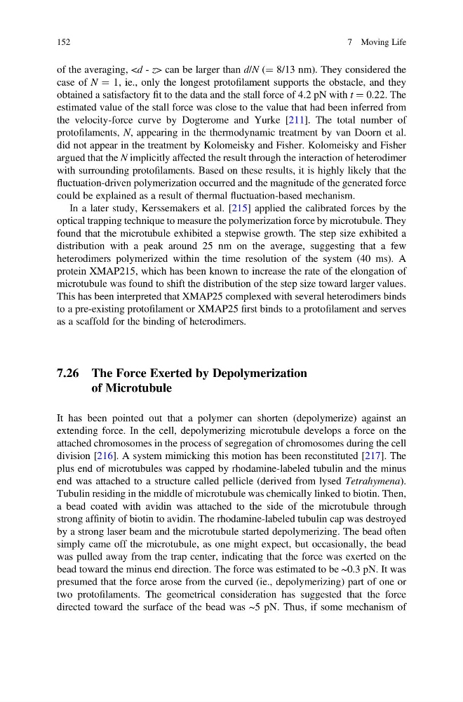
of the averaging, can be larger than d/N (¼ 8/13 nm). They considered the
case of N ¼ 1, ie., only the longest protofilament supports the obstacle, and they
obtained a satisfactory fit to the data and the stall force of 4.2 pN with t ¼ 0.22. The
estimated value of the stall force was close to the value that had been inferred from
the velocity-force curve by Dogterome and Yurke [211]. The total number of
protofilaments, N, appearing in the thermodynamic treatment by van Doorn et al.
did not appear in the treatment by Kolomeisky and Fisher. Kolomeisky and Fisher
argued that the N implicitly affected the result through the interaction of heterodimer
with surrounding protofilaments. Based on these results, it is highly likely that the
fluctuation-driven polymerization occurred and the magnitude of the generated force
could be explained as a result of thermal fluctuation-based mechanism.
In a later study, Kerssemakers et al. [215] applied the calibrated forces by the
optical trapping technique to measure the polymerization force by microtubule. They
found that the microtubule exhibited a stepwise growth. The step size exhibited a
distribution with a peak around 25 nm on the average, suggesting that a few
heterodimers polymerized within the time resolution of the system (40 ms). A
protein XMAP215, which has been known to increase the rate of the elongation of
microtubule was found to shift the distribution of the step size toward larger values.
This has been interpreted that XMAP25 complexed with several heterodimers binds
to a pre-existing protofilament or XMAP25 first binds to a protofilament and serves
as a scaffold for the binding of heterodimers.
7.26 The Force Exerted by Depolymerization
of Microtubule
It has been pointed out that a polymer can shorten (depolymerize) against an
extending force. In the cell, depolymerizing microtubule develops a force on the
attached chromosomes in the process of segregation of chromosomes during the cell
division [216]. A system mimicking this motion has been reconstituted [217]. The
plus end of microtubules was capped by rhodamine-labeled tubulin and the minus
end was attached to a structure called pellicle (derived from lysed Tetrahymena).
Tubulin residing in the middle of microtubule was chemically linked to biotin. Then,
a bead coated with avidin was attached to the side of the microtubule through
strong affinity of biotin to avidin. The rhodamine-labeled tubulin cap was destroyed
by a strong laser beam and the microtubule started depolymerizing. The bead often
simply came off the microtubule, as one might expect, but occasionally, the bead
was pulled away from the trap center, indicating that the force was exerted on the
bead toward the minus end direction. The force was estimated to be ~0.3 pN. It was
presumed that the force arose from the curved (ie., depolymerizing) part of one or
two protofilaments. The geometrical consideration has suggested that the force
directed toward the surface of the bead was ~5 pN. Thus, if some mechanism of
152
7 Moving Life
case of N ¼ 1, ie., only the longest protofilament supports the obstacle, and they
obtained a satisfactory fit to the data and the stall force of 4.2 pN with t ¼ 0.22. The
estimated value of the stall force was close to the value that had been inferred from
the velocity-force curve by Dogterome and Yurke [211]. The total number of
protofilaments, N, appearing in the thermodynamic treatment by van Doorn et al.
did not appear in the treatment by Kolomeisky and Fisher. Kolomeisky and Fisher
argued that the N implicitly affected the result through the interaction of heterodimer
with surrounding protofilaments. Based on these results, it is highly likely that the
fluctuation-driven polymerization occurred and the magnitude of the generated force
could be explained as a result of thermal fluctuation-based mechanism.
In a later study, Kerssemakers et al. [215] applied the calibrated forces by the
optical trapping technique to measure the polymerization force by microtubule. They
found that the microtubule exhibited a stepwise growth. The step size exhibited a
distribution with a peak around 25 nm on the average, suggesting that a few
heterodimers polymerized within the time resolution of the system (40 ms). A
protein XMAP215, which has been known to increase the rate of the elongation of
microtubule was found to shift the distribution of the step size toward larger values.
This has been interpreted that XMAP25 complexed with several heterodimers binds
to a pre-existing protofilament or XMAP25 first binds to a protofilament and serves
as a scaffold for the binding of heterodimers.
7.26 The Force Exerted by Depolymerization
of Microtubule
It has been pointed out that a polymer can shorten (depolymerize) against an
extending force. In the cell, depolymerizing microtubule develops a force on the
attached chromosomes in the process of segregation of chromosomes during the cell
division [216]. A system mimicking this motion has been reconstituted [217]. The
plus end of microtubules was capped by rhodamine-labeled tubulin and the minus
end was attached to a structure called pellicle (derived from lysed Tetrahymena).
Tubulin residing in the middle of microtubule was chemically linked to biotin. Then,
a bead coated with avidin was attached to the side of the microtubule through
strong affinity of biotin to avidin. The rhodamine-labeled tubulin cap was destroyed
by a strong laser beam and the microtubule started depolymerizing. The bead often
simply came off the microtubule, as one might expect, but occasionally, the bead
was pulled away from the trap center, indicating that the force was exerted on the
bead toward the minus end direction. The force was estimated to be ~0.3 pN. It was
presumed that the force arose from the curved (ie., depolymerizing) part of one or
two protofilaments. The geometrical consideration has suggested that the force
directed toward the surface of the bead was ~5 pN. Thus, if some mechanism of
152
7 Moving Life